 Movie
Movie Controller


+ Open data

- Basic information
| Entry | Database: PDB / ID: 7zzy | |||||||||||||||||||||||||||||||||||||||||||||||||||
|---|---|---|---|---|---|---|---|---|---|---|---|---|---|---|---|---|---|---|---|---|---|---|---|---|---|---|---|---|---|---|---|---|---|---|---|---|---|---|---|---|---|---|---|---|---|---|---|---|---|---|---|---|
| Title | Solution BcsD structure | |||||||||||||||||||||||||||||||||||||||||||||||||||
 Components Components | Cellulose biosynthesis protein | |||||||||||||||||||||||||||||||||||||||||||||||||||
 Keywords Keywords | STRUCTURAL PROTEIN / Bacterial cytoskeleton / Crystalline cellulose secretion | |||||||||||||||||||||||||||||||||||||||||||||||||||
| Function / homology | Cellulose synthase operon protein D, bacterial / Cellulose synthase subunit D superfamily / Cellulose synthase subunit D / cellulose biosynthetic process / Cellulose biosynthesis protein Function and homology information Function and homology information | |||||||||||||||||||||||||||||||||||||||||||||||||||
| Biological species |  Komagataeibacter hansenii ATCC 23769 (bacteria) Komagataeibacter hansenii ATCC 23769 (bacteria) | |||||||||||||||||||||||||||||||||||||||||||||||||||
| Method | ELECTRON MICROSCOPY / single particle reconstruction / cryo EM / Resolution: 3.3 Å | |||||||||||||||||||||||||||||||||||||||||||||||||||
 Authors Authors | Krasteva, P.V. / Abidi, W. / Decossas, M. | |||||||||||||||||||||||||||||||||||||||||||||||||||
| Funding support | European Union, 1items
| |||||||||||||||||||||||||||||||||||||||||||||||||||
 Citation Citation | Journal: Sci Adv / Year: 2022 Title: Bacterial crystalline cellulose secretion via a supramolecular BcsHD scaffold. Authors: Wiem Abidi / Marion Decossas / Lucía Torres-Sánchez / Lucie Puygrenier / Sylvie Létoffé / Jean-Marc Ghigo / Petya V Krasteva /  Abstract: Cellulose, the most abundant biopolymer on Earth, is not only the predominant constituent of plants but also a key extracellular polysaccharide in the biofilms of many bacterial species. Depending on ...Cellulose, the most abundant biopolymer on Earth, is not only the predominant constituent of plants but also a key extracellular polysaccharide in the biofilms of many bacterial species. Depending on the producers, chemical modifications, and three-dimensional assemblies, bacterial cellulose (BC) can present diverse degrees of crystallinity. Highly ordered, or crystalline, cellulose presents great economical relevance due to its ever-growing number of biotechnological applications. Even if some acetic acid bacteria have long been identified as BC superproducers, the molecular mechanisms determining the secretion of crystalline versus amorphous cellulose remain largely unknown. Here, we present structural and mechanistic insights into the role of the accessory subunits BcsH (CcpAx) and BcsD (CesD) that determine crystalline BC secretion in the lineage. We show that oligomeric BcsH drives the assembly of BcsD into a supramolecular cytoskeletal scaffold that likely stabilizes the cellulose-extruding synthase nanoarrays through an unexpected inside-out mechanism for secretion system assembly. | |||||||||||||||||||||||||||||||||||||||||||||||||||
| History |
|
- Structure visualization
| Structure viewer | Molecule: MolmilJmol/JSmol |
|---|
- Downloads & links
-Download
| PDBx/mmCIF format | 7zzy.cif.gz | 200.7 KB | Display | PDBx/mmCIF format |
|---|---|---|---|---|
| PDB format | pdb7zzy.ent.gz | 159.4 KB | Display | PDB format |
| PDBx/mmJSON format | 7zzy.json.gz | Tree view | PDBx/mmJSON format | |
| Others |  Other downloads Other downloads |
-Validation report
| Arichive directory | https://data.pdbj.org/pub/pdb/validation_reports/zz/7zzyftp://data.pdbj.org/pub/pdb/validation_reports/zz/7zzy | HTTPS FTP |
|---|
-Related structure data
| Related structure data |  15041MC  7zzqC C: citing same article ( M: map data used to model this data |
|---|---|
| Similar structure data |


-Links
 PDBj
PDBj- Assembly
| Deposited unit | 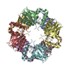
|
|---|---|
| 1 |
|
-Components
| #1: Protein | Mass: 20596.332 Da / Num. of mol.: 8 Source method: isolated from a genetically manipulated source Details: The HRV3c-cleavable hexahistidine tag was cleaved during the purification process. Sample protein sequence starts with PMGSTIFEK... Source: (gene. exp.) Komagataeibacter hansenii ATCC 23769 (bacteria)Gene: acsD, GXY_04292 / Plasmid: pProEx-BcsD / Production host: #2: Water | ChemComp-HOH / |  Mass: 18.015 Da / Num. of mol.: 24 / Source method: isolated from a natural source / Formula: H2O Mass: 18.015 Da / Num. of mol.: 24 / Source method: isolated from a natural source / Formula: H2OHas protein modification | N | |
|---|
-Experimental details
-Experiment
| Experiment | Method: ELECTRON MICROSCOPY |
|---|---|
| EM experiment | Aggregation state: PARTICLE / 3D reconstruction method: single particle reconstruction |
- Sample preparation
| Component | Name: BcsD from G. hansenii / Type: COMPLEX Details: Octameric BcsD in solution. Expressed recombinantly in E. coli and purified via a cleavable N-terminal hexahistidine tag. Entity ID: #1 / Source: RECOMBINANT |
|---|---|
| Molecular weight | Experimental value: NO |
| Source (natural) | Organism: Komagataeibacter hansenii ATCC 23769 (bacteria) / Strain: ATCC 23769 |
| Source (recombinant) | Organism: |
| Buffer solution | pH: 8 / Details: 20 mM HEPES pH 8.0, 100 mM NaCl |
| Specimen | Conc.: 1.8 mg/ml / Embedding applied: NO / Shadowing applied: NO / Staining applied: NO / Vitrification applied: YES |
| Specimen support | Grid material: GOLD / Grid mesh size: 300 divisions/in. / Grid type: Quantifoil R1.2/1.3 |
| Vitrification | Instrument: FEI VITROBOT MARK IV / Cryogen name: ETHANE |
- Electron microscopy imaging
| Experimental equipment |  Model: Talos Arctica / Image courtesy: FEI Company |
|---|---|
| Microscopy | Model: FEI TALOS ARCTICA |
| Electron gun | Electron source:  FIELD EMISSION GUN / Accelerating voltage: 200 kV / Illumination mode: FLOOD BEAM FIELD EMISSION GUN / Accelerating voltage: 200 kV / Illumination mode: FLOOD BEAM |
| Electron lens | Mode: BRIGHT FIELD / Nominal defocus max: 2750 nm / Nominal defocus min: 480 nm / Cs: 2.7 mm |
| Image recording | Electron dose: 40.59 e/Å2 / Detector mode: COUNTING / Film or detector model: GATAN K2 SUMMIT (4k x 4k) |
- Processing
| Software | Name: PHENIX / Version: 1.19.2_4158: / Classification: refinement | ||||||||||||||||||||||||
|---|---|---|---|---|---|---|---|---|---|---|---|---|---|---|---|---|---|---|---|---|---|---|---|---|---|
| EM software |
| ||||||||||||||||||||||||
| CTF correction | Type: PHASE FLIPPING AND AMPLITUDE CORRECTION | ||||||||||||||||||||||||
| Particle selection | Num. of particles selected: 1221383 | ||||||||||||||||||||||||
| Symmetry | Point symmetry: D4 (2x4 fold dihedral) | ||||||||||||||||||||||||
| 3D reconstruction | Resolution: 3.3 Å / Resolution method: FSC 0.143 CUT-OFF / Num. of particles: 380318 / Symmetry type: POINT | ||||||||||||||||||||||||
| Refine LS restraints |
|
