 Movie
Movie Controller

[English] 日本語
 Yorodumi
- PDB-7zyv: Cryo-EM structure of catalytically active Spinacia oleracea cytoc... -
+ Open data

- Basic information
| Entry | Database: PDB / ID: 7zyv | |||||||||
|---|---|---|---|---|---|---|---|---|---|---|
| Title | Cryo-EM structure of catalytically active Spinacia oleracea cytochrome b6f in complex with endogenous plastoquinones at 2.13 A resolution | |||||||||
 Components Components |
| |||||||||
 Keywords Keywords | PHOTOSYNTHESIS / cytochrome / Spinacia oleracea / plastoquinones / cryo-EM | |||||||||
| Function / homology |  Function and homology information Function and homology informationPSII associated light-harvesting complex II binding / chloroplast photosystem I binding / chloroplast photosystem II binding / plastoquinol--plastocyanin reductase activity / cytochrome b6f complex / plastoquinol-plastocyanin reductase / electron transporter, transferring electrons within cytochrome b6/f complex of photosystem II activity / thylakoid membrane / cytochrome complex assembly / : ...PSII associated light-harvesting complex II binding / chloroplast photosystem I binding / chloroplast photosystem II binding / plastoquinol--plastocyanin reductase activity / cytochrome b6f complex / plastoquinol-plastocyanin reductase / electron transporter, transferring electrons within cytochrome b6/f complex of photosystem II activity / thylakoid membrane / cytochrome complex assembly / : / photosynthetic electron transport chain / ubiquinol-cytochrome-c reductase activity / mitochondrial electron transport, ubiquinol to cytochrome c / electron transporter, transferring electrons within the cyclic electron transport pathway of photosynthesis activity / chloroplast thylakoid membrane / response to light stimulus / localization / photosynthesis / chloroplast / 2 iron, 2 sulfur cluster binding / electron transfer activity / iron ion binding / lipid binding / heme binding / membrane / metal ion binding / plasma membrane Similarity search - Function | |||||||||
| Biological species |  Spinacia oleracea (spinach) Spinacia oleracea (spinach) | |||||||||
| Method | ELECTRON MICROSCOPY / single particle reconstruction / cryo EM / Resolution: 2.13 Å | |||||||||
 Authors Authors | Sarewicz, M. / Szwalec, M. / Pintscher, S. / Indyka, P. / Rawski, M. / Pietras, R. / Mielecki, B. / Koziej, L. / Jaciuk, M. / Glatt, S. / Osyczka, A. | |||||||||
| Funding support |  Poland, 2items Poland, 2items
| |||||||||
 Citation Citation | Journal: Sci Adv / Year: 2023 Title: High-resolution cryo-EM structures of plant cytochrome bf at work. Authors: Marcin Sarewicz / Mateusz Szwalec / Sebastian Pintscher / Paulina Indyka / Michał Rawski / Rafał Pietras / Bohun Mielecki / Łukasz Koziej / Marcin Jaciuk / Sebastian Glatt / Artur Osyczka / Abstract: Plants use solar energy to power cellular metabolism. The oxidation of plastoquinol and reduction of plastocyanin by cytochrome bf (Cyt bf) is known as one of the key steps of photosynthesis, but the ...Plants use solar energy to power cellular metabolism. The oxidation of plastoquinol and reduction of plastocyanin by cytochrome bf (Cyt bf) is known as one of the key steps of photosynthesis, but the catalytic mechanism in the plastoquinone oxidation site (Q) remains elusive. Here, we describe two high-resolution cryo-EM structures of the spinach Cyt bf homodimer with endogenous plastoquinones and in complex with plastocyanin. Three plastoquinones are visible and line up one after another head to tail near Q in both monomers, indicating the existence of a channel in each monomer. Therefore, quinones appear to flow through Cyt bf in one direction, transiently exposing the redox-active ring of quinone during catalysis. Our work proposes an unprecedented one-way traffic model that explains efficient quinol oxidation during photosynthesis and respiration. | |||||||||
| History |
|
- Structure visualization
| Structure viewer | Molecule: MolmilJmol/JSmol |
|---|
- Downloads & links
-Download
| PDBx/mmCIF format | 7zyv.cif.gz | 383.4 KB | Display | PDBx/mmCIF format |
|---|---|---|---|---|
| PDB format | pdb7zyv.ent.gz | 318.3 KB | Display | PDB format |
| PDBx/mmJSON format | 7zyv.json.gz | Tree view | PDBx/mmJSON format | |
| Others |  Other downloads Other downloads |
-Validation report
| Summary document | 7zyv_validation.pdf.gz | 2.4 MB | Display | wwPDB validaton report |
|---|---|---|---|---|
| Full document | 7zyv_full_validation.pdf.gz | 2.5 MB | Display | |
| Data in XML | 7zyv_validation.xml.gz | 84.6 KB | Display | |
| Data in CIF | 7zyv_validation.cif.gz | 115.9 KB | Display | |
| Arichive directory | https://data.pdbj.org/pub/pdb/validation_reports/zy/7zyvftp://data.pdbj.org/pub/pdb/validation_reports/zy/7zyv | HTTPS FTP |
-Related structure data
| Related structure data | 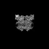 15027MC  7qrmC C: citing same article ( M: map data used to model this data |
|---|---|
| Similar structure data |

-Links
 PDBj
PDBj


- Assembly
| Deposited unit | 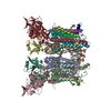
|
|---|---|
| 1 |
|
-Components
-Protein , 4 types, 8 molecules AICKDLRQ
| #1: Protein | Mass: 24186.504 Da / Num. of mol.: 2 / Source method: isolated from a natural source / Source: (natural) Spinacia oleracea (spinach) / References: UniProt: P00165#3: Protein | Mass: 35355.664 Da / Num. of mol.: 2 / Source method: isolated from a natural source / Source: (natural) Spinacia oleracea (spinach) / References: UniProt: P16013#4: Protein | Mass: 24347.898 Da / Num. of mol.: 2 / Source method: isolated from a natural source / Source: (natural) Spinacia oleracea (spinach)References: UniProt: P08980, plastoquinol-plastocyanin reductase #9: Protein | Mass: 10608.004 Da / Num. of mol.: 2 / Source method: isolated from a natural source / Source: (natural) Spinacia oleracea (spinach) / References: UniProt: Q8GT36 |
|---|
-Cytochrome b6-f complex subunit ... , 5 types, 10 molecules BJEMFNGOHP
| #2: Protein | Mass: 17456.662 Da / Num. of mol.: 2 / Source method: isolated from a natural source / Source: (natural) Spinacia oleracea (spinach) / References: UniProt: P00166#5: Protein/peptide | Mass: 3452.200 Da / Num. of mol.: 2 / Source method: isolated from a natural source / Source: (natural) Spinacia oleracea (spinach) / References: UniProt: Q9M3L0#6: Protein | Mass: 12953.800 Da / Num. of mol.: 2 / Source method: isolated from a natural source / Source: (natural) Spinacia oleracea (spinach) / References: UniProt: A0A0K9RMM1#7: Protein/peptide | Mass: 4171.985 Da / Num. of mol.: 2 / Source method: isolated from a natural source / Source: (natural) Spinacia oleracea (spinach) / References: UniProt: P69461#8: Protein/peptide | Mass: 3170.809 Da / Num. of mol.: 2 / Source method: isolated from a natural source / Source: (natural) Spinacia oleracea (spinach) / References: UniProt: P61045 |
|---|
-Non-polymers , 8 types, 32 molecules 


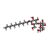
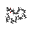


| #10: Chemical | ChemComp-HEM /  Mass: 616.487 Da / Num. of mol.: 4 / Source method: obtained synthetically / Formula: C34H32FeN4O4 / Feature type: SUBJECT OF INVESTIGATION Mass: 616.487 Da / Num. of mol.: 4 / Source method: obtained synthetically / Formula: C34H32FeN4O4 / Feature type: SUBJECT OF INVESTIGATION#11: Chemical | ChemComp-HEC /  Mass: 618.503 Da / Num. of mol.: 4 / Source method: obtained synthetically / Formula: C34H34FeN4O4 / Feature type: SUBJECT OF INVESTIGATION Mass: 618.503 Da / Num. of mol.: 4 / Source method: obtained synthetically / Formula: C34H34FeN4O4 / Feature type: SUBJECT OF INVESTIGATION#12: Chemical |  Mass: 893.489 Da / Num. of mol.: 2 / Source method: obtained synthetically / Formula: C55H72MgN4O5 / Feature type: SUBJECT OF INVESTIGATION Mass: 893.489 Da / Num. of mol.: 2 / Source method: obtained synthetically / Formula: C55H72MgN4O5 / Feature type: SUBJECT OF INVESTIGATION#13: Chemical | ChemComp-UMQ /  Mass: 496.589 Da / Num. of mol.: 10 / Source method: obtained synthetically / Formula: C23H44O11 / Feature type: SUBJECT OF INVESTIGATION / Comment: detergent*YM Mass: 496.589 Da / Num. of mol.: 10 / Source method: obtained synthetically / Formula: C23H44O11 / Feature type: SUBJECT OF INVESTIGATION / Comment: detergent*YM#14: Chemical | ChemComp-PL9 /  Mass: 749.201 Da / Num. of mol.: 6 / Source method: obtained synthetically / Formula: C53H80O2 / Feature type: SUBJECT OF INVESTIGATION Mass: 749.201 Da / Num. of mol.: 6 / Source method: obtained synthetically / Formula: C53H80O2 / Feature type: SUBJECT OF INVESTIGATION#15: Chemical |  Mass: 795.116 Da / Num. of mol.: 2 / Source method: obtained synthetically / Formula: C41H78O12S / Feature type: SUBJECT OF INVESTIGATION Mass: 795.116 Da / Num. of mol.: 2 / Source method: obtained synthetically / Formula: C41H78O12S / Feature type: SUBJECT OF INVESTIGATION#16: Chemical |  Mass: 175.820 Da / Num. of mol.: 2 / Source method: obtained synthetically / Formula: Fe2S2 / Feature type: SUBJECT OF INVESTIGATION Mass: 175.820 Da / Num. of mol.: 2 / Source method: obtained synthetically / Formula: Fe2S2 / Feature type: SUBJECT OF INVESTIGATION#17: Chemical |  Mass: 536.873 Da / Num. of mol.: 2 / Source method: obtained synthetically / Formula: C40H56 / Feature type: SUBJECT OF INVESTIGATION Mass: 536.873 Da / Num. of mol.: 2 / Source method: obtained synthetically / Formula: C40H56 / Feature type: SUBJECT OF INVESTIGATION |
|---|
-Details
| Has ligand of interest | Y |
|---|
-Experimental details
-Experiment
| Experiment | Method: ELECTRON MICROSCOPY |
|---|---|
| EM experiment | Aggregation state: PARTICLE / 3D reconstruction method: single particle reconstruction |
- Sample preparation
| Component | Name: Cytochrome b6f complex / Type: COMPLEX / Entity ID: #1-#9 / Source: NATURAL | ||||||||||||||||||||
|---|---|---|---|---|---|---|---|---|---|---|---|---|---|---|---|---|---|---|---|---|---|
| Molecular weight | Value: 0.22 MDa / Experimental value: NO | ||||||||||||||||||||
| Source (natural) | Organism: Spinacia oleracea (spinach) / Cellular location: thylakoids / Organ: Leaves / Organelle: Chloroplasts | ||||||||||||||||||||
| Buffer solution | pH: 8 Details: Buffer and the diluted protein sample were filtered through a 0.22 um syringe filter. Prior to freezing, protein sample was concentrated on centrifugal filter units with 50K MWCO. | ||||||||||||||||||||
| Buffer component |
| ||||||||||||||||||||
| Specimen | Conc.: 6.2 mg/ml / Embedding applied: NO / Shadowing applied: NO / Staining applied: NO / Vitrification applied: YES | ||||||||||||||||||||
| Specimen support | Grid material: COPPER / Grid mesh size: 200 divisions/in. / Grid type: Quantifoil R2/1 | ||||||||||||||||||||
| Vitrification | Instrument: FEI VITROBOT MARK IV / Cryogen name: ETHANE / Humidity: 100 % / Chamber temperature: 277 K / Details: blot time 2 s, wait time 0 s, blot force -1 |
- Electron microscopy imaging
| Experimental equipment |  Model: Titan Krios / Image courtesy: FEI Company |
|---|---|
| Microscopy | Model: TFS KRIOS |
| Electron gun | Electron source:  FIELD EMISSION GUN / Accelerating voltage: 300 kV / Illumination mode: FLOOD BEAM FIELD EMISSION GUN / Accelerating voltage: 300 kV / Illumination mode: FLOOD BEAM |
| Electron lens | Mode: BRIGHT FIELD / Nominal magnification: 105000 X / Nominal defocus max: 2100 nm / Nominal defocus min: 900 nm / Cs: 2.7 mm / C2 aperture diameter: 50 µm / Alignment procedure: COMA FREE |
| Specimen holder | Cryogen: NITROGEN / Specimen holder model: FEI TITAN KRIOS AUTOGRID HOLDER |
| Image recording | Average exposure time: 1.82 sec. / Electron dose: 40 e/Å2 / Film or detector model: GATAN K3 BIOQUANTUM (6k x 4k) / Num. of real images: 7651 |
| EM imaging optics | Energyfilter name: GIF Bioquantum / Energyfilter slit width: 20 eV |
| Image scans | Width: 5760 / Height: 4092 |
- Processing
| EM software |
| ||||||||||||||||||||||||||||||||||||||||||||||||||
|---|---|---|---|---|---|---|---|---|---|---|---|---|---|---|---|---|---|---|---|---|---|---|---|---|---|---|---|---|---|---|---|---|---|---|---|---|---|---|---|---|---|---|---|---|---|---|---|---|---|---|---|
| Image processing | Details: 20 eV slit, fully tuned before the experiment | ||||||||||||||||||||||||||||||||||||||||||||||||||
| CTF correction | Details: Non-uniform Refinement with iterative global CTF refinement and anisotropic magnification fitting Type: PHASE FLIPPING AND AMPLITUDE CORRECTION | ||||||||||||||||||||||||||||||||||||||||||||||||||
| Particle selection | Num. of particles selected: 42951 Details: given number of particles is after blob picker and 2D cleaning | ||||||||||||||||||||||||||||||||||||||||||||||||||
| Symmetry | Point symmetry: C2 (2 fold cyclic) | ||||||||||||||||||||||||||||||||||||||||||||||||||
| 3D reconstruction | Resolution: 2.13 Å / Resolution method: FSC 0.143 CUT-OFF / Num. of particles: 685979 / Details: Non-uniform Refinement / Symmetry type: POINT | ||||||||||||||||||||||||||||||||||||||||||||||||||
| Atomic model building | Protocol: RIGID BODY FIT | ||||||||||||||||||||||||||||||||||||||||||||||||||
| Atomic model building | PDB-ID: 7QRM |
