 Movie
Movie Controller

[English] 日本語
 Yorodumi
- PDB-7zxb: Crystal structure of human STING in complex with 3',3'-c-(2'dAMP-... -
+ Open data

- Basic information
| Entry | Database: PDB / ID: 7zxb | ||||||||||||||||||||||||||||||
|---|---|---|---|---|---|---|---|---|---|---|---|---|---|---|---|---|---|---|---|---|---|---|---|---|---|---|---|---|---|---|---|
| Title | Crystal structure of human STING in complex with 3',3'-c-(2'dAMP-2'F,2'd Components ComponentsStimulator of interferon protein |  Keywords KeywordsANTIVIRAL PROTEIN / sting / antiviral / activator | Function / homology |  Function and homology information Function and homology informationautophagosome membrane / positive regulation of type I interferon production / endoplasmic reticulum-Golgi intermediate compartment membrane / activation of innate immune response / mitochondrial outer membrane / nucleotide binding / endoplasmic reticulum membrane / perinuclear region of cytoplasm Similarity search - Function Biological species |  Homo sapiens (human) Homo sapiens (human)Method |  X-RAY DIFFRACTION / SYNCHROTRON / MOLECULAR REPLACEMENT / Resolution: 3 Å X-RAY DIFFRACTION / SYNCHROTRON / MOLECULAR REPLACEMENT / Resolution: 3 Å  Authors AuthorsKlima, M. / Smola, M. / Boura, E. | Funding support | 1items |
 CitationJournal: To Be Published CitationJournal: To Be PublishedTitle: Crystal structure of human STING in complex with 3',3'-c-(2'dAMP-2'F,2'd Authors: Klima, M. / Smola, M. / Boura, E. History |
|
- Structure visualization
| Structure viewer | Molecule: MolmilJmol/JSmol |
|---|
- Downloads & links
-Download
| PDBx/mmCIF format | 7zxb.cif.gz | 53.2 KB | Display | PDBx/mmCIF format |
|---|---|---|---|---|
| PDB format | pdb7zxb.ent.gz | 34.3 KB | Display | PDB format |
| PDBx/mmJSON format | 7zxb.json.gz | Tree view | PDBx/mmJSON format | |
| Others |  Other downloads Other downloads |
-Validation report
| Summary document | 7zxb_validation.pdf.gz | 859.1 KB | Display | wwPDB validaton report |
|---|---|---|---|---|
| Full document | 7zxb_full_validation.pdf.gz | 859.1 KB | Display | |
| Data in XML | 7zxb_validation.xml.gz | 8 KB | Display | |
| Data in CIF | 7zxb_validation.cif.gz | 9.9 KB | Display | |
| Arichive directory | https://data.pdbj.org/pub/pdb/validation_reports/zx/7zxbftp://data.pdbj.org/pub/pdb/validation_reports/zx/7zxb | HTTPS FTP |
-Related structure data
| Related structure data |  4ksyS S: Starting model for refinement |
|---|---|
| Similar structure data |
-Links
 PDBj
PDBj- Assembly
| Deposited unit | 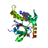
| ||||||||||||
|---|---|---|---|---|---|---|---|---|---|---|---|---|---|
| 1 | 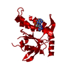
| ||||||||||||
| Unit cell |
|
-Components
| #1: Protein | Mass: 23189.064 Da / Num. of mol.: 1 Source method: isolated from a genetically manipulated source Source: (gene. exp.) Homo sapiens (human) / Gene: STING, LOC340061, hCG_1782396 / Production host:  |
|---|---|
| #2: Chemical | ChemComp-KAX /   Mass: 642.431 Da / Num. of mol.: 1 / Source method: obtained synthetically / Formula: C21H25FN10O9P2 / Feature type: SUBJECT OF INVESTIGATION Mass: 642.431 Da / Num. of mol.: 1 / Source method: obtained synthetically / Formula: C21H25FN10O9P2 / Feature type: SUBJECT OF INVESTIGATION |
| Has ligand of interest | Y |
-Experimental details
-Experiment
| Experiment | Method: X-RAY DIFFRACTION / Number of used crystals: 1 |
|---|
- Sample preparation
| Crystal | Density Matthews: 2.37 Å3/Da / Density % sol: 48.01 % |
|---|---|
| Crystal grow | Temperature: 291 K / Method: vapor diffusion, sitting drop / Details: 0.2 M Lithium acetate, 20% (w/v) PEG 3350 |
-Data collection
| Diffraction | Mean temperature: 100 K / Serial crystal experiment: N |
|---|---|
| Diffraction source | Source: SYNCHROTRON / Site: BESSY  / Beamline: 14.1 / Wavelength: 0.9184 Å / Beamline: 14.1 / Wavelength: 0.9184 Å |
| Detector | Type: DECTRIS PILATUS 6M / Detector: PIXEL / Date: Feb 11, 2019 |
| Radiation | Protocol: SINGLE WAVELENGTH / Monochromatic (M) / Laue (L): M / Scattering type: x-ray |
| Radiation wavelength | Wavelength: 0.9184 Å / Relative weight: 1 |
| Reflection | Resolution: 3→35.36 Å / Num. obs: 4804 / % possible obs: 99.61 % / Redundancy: 16.2 % / Biso Wilson estimate: 71.06 Å2 / CC1/2: 0.998 / CC star: 1 / Rmerge(I) obs: 0.1612 / Rpim(I) all: 0.04082 / Rrim(I) all: 0.1665 / Net I/σ(I): 17.29 |
| Reflection shell | Resolution: 3→3.107 Å / Redundancy: 17.3 % / Rmerge(I) obs: 1.256 / Mean I/σ(I) obs: 2.59 / Num. unique obs: 465 / CC1/2: 0.806 / CC star: 0.945 / Rpim(I) all: 0.3076 / Rrim(I) all: 1.294 / % possible all: 100 |
- Processing
| Software |
| ||||||||||||||||||||||||||||
|---|---|---|---|---|---|---|---|---|---|---|---|---|---|---|---|---|---|---|---|---|---|---|---|---|---|---|---|---|---|
| Refinement | Method to determine structure: MOLECULAR REPLACEMENT Starting model: 4ksy Resolution: 3→35.36 Å / SU ML: 0.2088 / Cross valid method: FREE R-VALUE / σ(F): 1.35 / Phase error: 28.8023 Stereochemistry target values: GeoStd + Monomer Library + CDL v1.2
| ||||||||||||||||||||||||||||
| Solvent computation | Shrinkage radii: 0.9 Å / VDW probe radii: 1.1 Å / Solvent model: FLAT BULK SOLVENT MODEL | ||||||||||||||||||||||||||||
| Displacement parameters | Biso mean: 63.3 Å2 | ||||||||||||||||||||||||||||
| Refinement step | Cycle: LAST / Resolution: 3→35.36 Å
| ||||||||||||||||||||||||||||
| Refine LS restraints |
| ||||||||||||||||||||||||||||
| LS refinement shell |
|
