 Movie
Movie Controller

[English] 日本語
 Yorodumi
- PDB-7zv0: Crystal structure of human STING in complex with 3',3'-c-(2'F,2'd... -
+ Open data

- Basic information
| Entry | Database: PDB / ID: 7zv0 | ||||||||||||||||||||||||||||||
|---|---|---|---|---|---|---|---|---|---|---|---|---|---|---|---|---|---|---|---|---|---|---|---|---|---|---|---|---|---|---|---|
| Title | Crystal structure of human STING in complex with 3',3'-c-(2'F,2'dAMP-2'F,2'd Components ComponentsStimulator of interferon protein |  Keywords KeywordsANTIVIRAL PROTEIN / sting / antiviral / activator | Function / homology |  Function and homology information Function and homology information2',3'-cyclic GMP-AMP binding / autophagosome membrane / positive regulation of type I interferon production / endoplasmic reticulum-Golgi intermediate compartment membrane / activation of innate immune response / mitochondrial outer membrane / Golgi membrane / endoplasmic reticulum membrane / perinuclear region of cytoplasm Similarity search - Function Biological species |  Homo sapiens (human) Homo sapiens (human)Method |  X-RAY DIFFRACTION / MOLECULAR REPLACEMENT / Resolution: 2.31 Å X-RAY DIFFRACTION / MOLECULAR REPLACEMENT / Resolution: 2.31 Å  Authors AuthorsKlima, M. / Smola, M. / Boura, E. | Funding support | 1items |
 CitationJournal: To Be Published CitationJournal: To Be PublishedTitle: Crystal structure of human STING in complex with 3',3'-c-(2'F,2'dAMP-2'F,2'd Authors: Klima, M. / Smola, M. / Boura, E. History |
|
- Structure visualization
| Structure viewer | Molecule: MolmilJmol/JSmol |
|---|
- Downloads & links
-Download
| PDBx/mmCIF format | 7zv0.cif.gz | 52.9 KB | Display | PDBx/mmCIF format |
|---|---|---|---|---|
| PDB format | pdb7zv0.ent.gz | 35.2 KB | Display | PDB format |
| PDBx/mmJSON format | 7zv0.json.gz | Tree view | PDBx/mmJSON format | |
| Others |  Other downloads Other downloads |
-Validation report
| Arichive directory | https://data.pdbj.org/pub/pdb/validation_reports/zv/7zv0ftp://data.pdbj.org/pub/pdb/validation_reports/zv/7zv0 | HTTPS FTP |
|---|
-Related structure data
| Related structure data |  4ksyS S: Starting model for refinement |
|---|---|
| Similar structure data |
-Links
 PDBj
PDBj- Assembly
| Deposited unit | 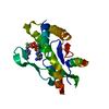
| ||||||||||||
|---|---|---|---|---|---|---|---|---|---|---|---|---|---|
| 1 | 
| ||||||||||||
| Unit cell |
| ||||||||||||
| Components on special symmetry positions |
|
-Components
| #1: Protein | Mass: 23189.064 Da / Num. of mol.: 1 Source method: isolated from a genetically manipulated source Source: (gene. exp.) Homo sapiens (human) / Gene: STING, LOC340061, hCG_1782396 / Production host:  |
|---|---|
| #2: Chemical | ChemComp-K1C / 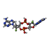  Mass: 660.421 Da / Num. of mol.: 1 / Source method: obtained synthetically / Formula: C21H24F2N10O9P2 / Feature type: SUBJECT OF INVESTIGATION Mass: 660.421 Da / Num. of mol.: 1 / Source method: obtained synthetically / Formula: C21H24F2N10O9P2 / Feature type: SUBJECT OF INVESTIGATION |
| #3: Water | ChemComp-HOH /  Mass: 18.015 Da / Num. of mol.: 59 / Source method: isolated from a natural source / Formula: H2O Mass: 18.015 Da / Num. of mol.: 59 / Source method: isolated from a natural source / Formula: H2O |
| Has ligand of interest | Y |
| Has protein modification | N |
-Experimental details
-Experiment
| Experiment | Method: X-RAY DIFFRACTION / Number of used crystals: 1 |
|---|
- Sample preparation
| Crystal | Density Matthews: 2.38 Å3/Da / Density % sol: 48.22 % |
|---|---|
| Crystal grow | Temperature: 291 K / Method: vapor diffusion, sitting drop / Details: 0.2 M Lithium acetate, 20% (w/v) PEG 3350 |
-Data collection
| Diffraction | Mean temperature: 100 K / Serial crystal experiment: N |
|---|---|
| Diffraction source | Source: ROTATING ANODE / Type: RIGAKU MICROMAX-007 HF / Wavelength: 1.54187 Å |
| Detector | Type: DECTRIS PILATUS 200K / Detector: PIXEL / Date: Jul 10, 2019 |
| Radiation | Protocol: SINGLE WAVELENGTH / Monochromatic (M) / Laue (L): M / Scattering type: x-ray |
| Radiation wavelength | Wavelength: 1.54187 Å / Relative weight: 1 |
| Reflection | Resolution: 2.31→34.08 Å / Num. obs: 10263 / % possible obs: 99.56 % / Redundancy: 9.5 % / Biso Wilson estimate: 29.16 Å2 / CC1/2: 0.997 / CC star: 0.999 / Rmerge(I) obs: 0.1526 / Rpim(I) all: 0.05162 / Rrim(I) all: 0.1614 / Net I/σ(I): 14.08 |
| Reflection shell | Resolution: 2.31→2.393 Å / Redundancy: 7.7 % / Rmerge(I) obs: 0.816 / Mean I/σ(I) obs: 2.34 / Num. unique obs: 983 / CC1/2: 0.801 / CC star: 0.943 / Rpim(I) all: 0.3036 / Rrim(I) all: 0.8729 / % possible all: 97.04 |
- Processing
| Software |
| ||||||||||||||||||||||||||||||||||||||||||||||||||||||||
|---|---|---|---|---|---|---|---|---|---|---|---|---|---|---|---|---|---|---|---|---|---|---|---|---|---|---|---|---|---|---|---|---|---|---|---|---|---|---|---|---|---|---|---|---|---|---|---|---|---|---|---|---|---|---|---|---|---|
| Refinement | Method to determine structure: MOLECULAR REPLACEMENT Starting model: 4ksy Resolution: 2.31→34.08 Å / SU ML: 0.2559 / Cross valid method: FREE R-VALUE / σ(F): 1.35 / Phase error: 21.6663 Stereochemistry target values: GeoStd + Monomer Library + CDL v1.2
| ||||||||||||||||||||||||||||||||||||||||||||||||||||||||
| Solvent computation | Shrinkage radii: 0.9 Å / VDW probe radii: 1.1 Å / Solvent model: FLAT BULK SOLVENT MODEL | ||||||||||||||||||||||||||||||||||||||||||||||||||||||||
| Displacement parameters | Biso mean: 32.54 Å2 | ||||||||||||||||||||||||||||||||||||||||||||||||||||||||
| Refinement step | Cycle: LAST / Resolution: 2.31→34.08 Å
| ||||||||||||||||||||||||||||||||||||||||||||||||||||||||
| Refine LS restraints |
| ||||||||||||||||||||||||||||||||||||||||||||||||||||||||
| LS refinement shell |
|
