 Movie
Movie Controller


+ Open data

- Basic information
| Entry | Database: PDB / ID: 7zud | |||||||||
|---|---|---|---|---|---|---|---|---|---|---|
| Title | Crystal structure of HIV-1 capsid IP6-CPSF6 complex | |||||||||
 Components Components |
| |||||||||
 Keywords Keywords | VIRAL PROTEIN / Complex / cofactor / host-factor | |||||||||
| Function / homology |  Function and homology information Function and homology informationexon-exon junction complex binding / positive regulation of RNA export from nucleus / mRNA cleavage factor complex / interchromatin granule / co-transcriptional mRNA 3'-end processing, cleavage and polyadenylation pathway / perichromatin fibrils / Processing of Intronless Pre-mRNAs / mRNA alternative polyadenylation / mRNA cleavage and polyadenylation specificity factor complex / mRNA 3'-end processing ...exon-exon junction complex binding / positive regulation of RNA export from nucleus / mRNA cleavage factor complex / interchromatin granule / co-transcriptional mRNA 3'-end processing, cleavage and polyadenylation pathway / perichromatin fibrils / Processing of Intronless Pre-mRNAs / mRNA alternative polyadenylation / mRNA cleavage and polyadenylation specificity factor complex / mRNA 3'-end processing / Signaling by cytosolic FGFR1 fusion mutants / mRNA 3'-end processing / paraspeckles / RNA Polymerase II Transcription Termination / protein heterotetramerization / ribosomal large subunit binding / Processing of Capped Intron-Containing Pre-mRNA / Signaling by FGFR1 in disease / HIV-1 retropepsin / symbiont-mediated activation of host apoptosis / protein tetramerization / retroviral ribonuclease H / exoribonuclease H / exoribonuclease H activity / DNA integration / viral genome integration into host DNA / establishment of integrated proviral latency / RNA-directed DNA polymerase / RNA stem-loop binding / viral penetration into host nucleus / host multivesicular body / RNA-directed DNA polymerase activity / mRNA processing / RNA-DNA hybrid ribonuclease activity / Transferases; Transferring phosphorus-containing groups; Nucleotidyltransferases / host cell / viral nucleocapsid / DNA recombination / DNA-directed DNA polymerase / aspartic-type endopeptidase activity / Hydrolases; Acting on ester bonds / DNA-directed DNA polymerase activity / nuclear speck / ribonucleoprotein complex / symbiont-mediated suppression of host gene expression / viral translational frameshifting / mRNA binding / symbiont entry into host cell / lipid binding / host cell nucleus / host cell plasma membrane / virion membrane / structural molecule activity / proteolysis / DNA binding / RNA binding / zinc ion binding / nucleoplasm / nucleus / membrane / cytoplasm Similarity search - Function | |||||||||
| Biological species |   Human immunodeficiency virus 1 Human immunodeficiency virus 1 Homo sapiens (human) Homo sapiens (human) | |||||||||
| Method |  X-RAY DIFFRACTION / SYNCHROTRON / MOLECULAR REPLACEMENT / Resolution: 2.93 Å X-RAY DIFFRACTION / SYNCHROTRON / MOLECULAR REPLACEMENT / Resolution: 2.93 Å | |||||||||
 Authors Authors | Nicastro, G. / Taylor, I.A. | |||||||||
| Funding support |  United Kingdom, 2items United Kingdom, 2items
| |||||||||
 Citation Citation | Journal: J.Mol.Biol. / Year: 2022 Title: CP-MAS and Solution NMR Studies of Allosteric Communication in CA-assemblies of HIV-1. Authors: Nicastro, G. / Lucci, M. / Oregioni, A. / Kelly, G. / Frenkiel, T.A. / Taylor, I.A. #1: Journal: Cell / Year: 2009Title: X-ray structures of the hexameric building block of the HIV capsid. Authors: Pornillos, O. / Ganser-Pornillos, B.K. / Kelly, B.N. / Hua, Y. / Whitby, F.G. / Stout, C.D. / Sundquist, W.I. / Hill, C.P. / Yeager, M. #2: Journal: Proc Natl Acad Sci U S A / Year: 2014Title: Structural basis of HIV-1 capsid recognition by PF74 and CPSF6. Authors: Bhattacharya, A. / Alam, S.L. / Fricke, T. / Zadrozny, K. / Sedzicki, J. / Taylor, A.B. / Demeler, B. / Pornillos, O. / Ganser-Pornillos, B.K. / Diaz-Griffero, F. / Ivanov, D.N. / Yeager, M. #3: Journal: PLoS Pathog / Year: 2014Title: Host cofactors and pharmacologic ligands share an essential interface in HIV-1 capsid that is lost upon disassembly. Authors: Price, A.J. / Jacques, D.A. / McEwan, W.A. / Fletcher, A.J. / Essig, S. / Chin, J.W. / Halambage, U.D. / Aiken, C. / James, L.C. #4: Journal: Nature / Year: 2018Title: Inositol phosphates are assembly co-factors for HIV-1. Authors: Dick, R.A. / Zadrozny, K.K. / Xu, C. / Schur, F.K.M. / Lyddon, T.D. / Ricana, C.L. / Wagner, J.M. / Perilla, J.R. / Ganser-Pornillos, B.K. / Johnson, M.C. / Pornillos, O. / Vogt, V.M. | |||||||||
| History |
|
- Structure visualization
| Structure viewer | Molecule: MolmilJmol/JSmol |
|---|
- Downloads & links
-Download
| PDBx/mmCIF format | 7zud.cif.gz | 128.2 KB | Display | PDBx/mmCIF format |
|---|---|---|---|---|
| PDB format | pdb7zud.ent.gz | 84.3 KB | Display | PDB format |
| PDBx/mmJSON format | 7zud.json.gz | Tree view | PDBx/mmJSON format | |
| Others |  Other downloads Other downloads |
-Validation report
| Arichive directory | https://data.pdbj.org/pub/pdb/validation_reports/zu/7zudftp://data.pdbj.org/pub/pdb/validation_reports/zu/7zud | HTTPS FTP |
|---|
-Related structure data
| Related structure data |  3h47S S: Starting model for refinement |
|---|---|
| Similar structure data |
-Links
 PDBj
PDBj


- Assembly
| Deposited unit | 
| |||||||||||||||
|---|---|---|---|---|---|---|---|---|---|---|---|---|---|---|---|---|
| 1 | x 6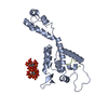
| |||||||||||||||
| Unit cell |
| |||||||||||||||
| Components on special symmetry positions |
|
-Components
| #1: Protein | Mass: 25703.545 Da / Num. of mol.: 1 Source method: isolated from a genetically manipulated source Source: (gene. exp.) Human immunodeficiency virus 1 / Strain: isolate NY5 / Gene: gag-pol / Production host:  | ||||||
|---|---|---|---|---|---|---|---|
| #2: Protein/peptide | Mass: 1380.587 Da / Num. of mol.: 1 / Source method: obtained synthetically / Source: (synth.) Homo sapiens (human) / References: UniProt: Q16630 | ||||||
| #3: Chemical |   Mass: 660.035 Da / Num. of mol.: 2 / Source method: obtained synthetically / Formula: C6H18O24P6 / Feature type: SUBJECT OF INVESTIGATION Mass: 660.035 Da / Num. of mol.: 2 / Source method: obtained synthetically / Formula: C6H18O24P6 / Feature type: SUBJECT OF INVESTIGATION#4: Water | ChemComp-HOH / |  Mass: 18.015 Da / Num. of mol.: 6 / Source method: isolated from a natural source / Formula: H2O Mass: 18.015 Da / Num. of mol.: 6 / Source method: isolated from a natural source / Formula: H2OHas ligand of interest | Y | Has protein modification | Y | |
-Experimental details
-Experiment
| Experiment | Method: X-RAY DIFFRACTION / Number of used crystals: 1 |
|---|
- Sample preparation
| Crystal | Density Matthews: 2.62 Å3/Da / Density % sol: 52.98 % |
|---|---|
| Crystal grow | Temperature: 291 K / Method: vapor diffusion, sitting drop Details: 0.3M Calcium chloride dihydrate, 0.3 M Magnesium chloride hexahydrate, 0.1 M Tris hydrochloride pH 8.5, 0.1 M Bicine, 20% PEG 4000 |
-Data collection
| Diffraction | Mean temperature: 100 K / Serial crystal experiment: N |
|---|---|
| Diffraction source | Source: SYNCHROTRON / Site: Diamond / Beamline: I24 / Wavelength: 0.9688 Å |
| Detector | Type: DECTRIS EIGER2 XE 16M / Detector: PIXEL / Date: May 17, 2019 |
| Radiation | Protocol: SINGLE WAVELENGTH / Monochromatic (M) / Laue (L): M / Scattering type: x-ray |
| Radiation wavelength | Wavelength: 0.9688 Å / Relative weight: 1 |
| Reflection | Resolution: 2.93→78.58 Å / Num. obs: 5698 / % possible obs: 98.4 % / Redundancy: 1 % / Biso Wilson estimate: 61.42 Å2 / CC1/2: 0.984 / Net I/σ(I): 7.6 |
| Reflection shell | Resolution: 2.93→3.23 Å / Num. unique obs: 1717 / CC1/2: 0.425 |
- Processing
| Software |
| ||||||||||||||||||||||||||||||||||||||||
|---|---|---|---|---|---|---|---|---|---|---|---|---|---|---|---|---|---|---|---|---|---|---|---|---|---|---|---|---|---|---|---|---|---|---|---|---|---|---|---|---|---|
| Refinement | Method to determine structure: MOLECULAR REPLACEMENT Starting model: 3h47 Resolution: 2.93→78.58 Å / SU ML: 0.5208 / Cross valid method: FREE R-VALUE / σ(F): 1.39 / Phase error: 39.819 Stereochemistry target values: GeoStd + Monomer Library + CDL v1.2
| ||||||||||||||||||||||||||||||||||||||||
| Solvent computation | Shrinkage radii: 0.9 Å / VDW probe radii: 1.11 Å / Solvent model: FLAT BULK SOLVENT MODEL | ||||||||||||||||||||||||||||||||||||||||
| Displacement parameters | Biso mean: 69.9 Å2 | ||||||||||||||||||||||||||||||||||||||||
| Refinement step | Cycle: LAST / Resolution: 2.93→78.58 Å
| ||||||||||||||||||||||||||||||||||||||||
| Refine LS restraints |
| ||||||||||||||||||||||||||||||||||||||||
| LS refinement shell |
| ||||||||||||||||||||||||||||||||||||||||
| Refinement TLS params. | Method: refined / Origin x: 67.231075919 Å / Origin y: 88.3320904565 Å / Origin z: 11.0531490963 Å
| ||||||||||||||||||||||||||||||||||||||||
| Refinement TLS group | Selection details: all |
