 Movie
Movie Controller

[English] 日本語
 Yorodumi
- PDB-7zti: F61V Cytochrome c prime beta from Methylococcus capsulatus (Bath)... -
+ Open data

- Basic information
| Entry | Database: PDB / ID: 7zti | ||||||
|---|---|---|---|---|---|---|---|
| Title | F61V Cytochrome c prime beta from Methylococcus capsulatus (Bath): CO Complex | ||||||
 Components Components | Cytochrome c | ||||||
 Keywords Keywords | OXIDOREDUCTASE / Cytochrome c prime / Beta-sheet / CO complex | ||||||
| Function / homology | Cytochrome P460 / Cytochrome P460 superfamily / Cytochrome P460 / metal ion binding / CARBON MONOXIDE / HEME C / Cytochrome c Function and homology information Function and homology information | ||||||
| Biological species |  Methylococcus capsulatus str. Bath (bacteria) Methylococcus capsulatus str. Bath (bacteria) | ||||||
| Method |  X-RAY DIFFRACTION / SYNCHROTRON / MOLECULAR REPLACEMENT / Resolution: 2.09 Å X-RAY DIFFRACTION / SYNCHROTRON / MOLECULAR REPLACEMENT / Resolution: 2.09 Å | ||||||
 Authors Authors | Adams, H.R. / Hough, M.A. | ||||||
| Funding support | 1items
| ||||||
 Citation Citation | Journal: J.Biol.Chem. / Year: 2023 Title: A heme pocket aromatic quadrupole modulates gas binding to cytochrome c'-beta : Implications for NO sensors. Authors: Adams, H.R. / Svistunenko, D.A. / Wilson, M.T. / Fujii, S. / Strange, R.W. / Hardy, Z.A. / Vazquez, P.A. / Dabritz, T. / Streblow, G.J. / Andrew, C.R. / Hough, M.A. | ||||||
| History |
|
- Structure visualization
| Structure viewer | Molecule: MolmilJmol/JSmol |
|---|
- Downloads & links
-Download
| PDBx/mmCIF format | 7zti.cif.gz | 75.3 KB | Display | PDBx/mmCIF format |
|---|---|---|---|---|
| PDB format | pdb7zti.ent.gz | 54.5 KB | Display | PDB format |
| PDBx/mmJSON format | 7zti.json.gz | Tree view | PDBx/mmJSON format | |
| Others |  Other downloads Other downloads |
-Validation report
| Arichive directory | https://data.pdbj.org/pub/pdb/validation_reports/zt/7ztiftp://data.pdbj.org/pub/pdb/validation_reports/zt/7zti | HTTPS FTP |
|---|
-Related structure data
| Related structure data | 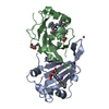 7zpsC 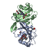 7zqzC  7zrwC 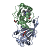 7zrxC  7zs4C 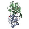 7zsvC  7zswC 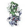 7zsxC  7zvzC  6hihS C: citing same article ( S: Starting model for refinement |
|---|---|
| Similar structure data |
-Links
 PDBj
PDBj


- Assembly
| Deposited unit | 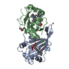
| ||||||||
|---|---|---|---|---|---|---|---|---|---|
| 1 |
| ||||||||
| Unit cell |
| ||||||||
| Components on special symmetry positions |
|
-Components
-Protein , 1 types, 2 molecules AB
| #1: Protein | Mass: 17322.607 Da / Num. of mol.: 2 Source method: isolated from a genetically manipulated source Source: (gene. exp.) Methylococcus capsulatus str. Bath (bacteria)Gene: ccp, MCA2394 / Production host: |
|---|
-Non-polymers , 5 types, 85 molecules 


| #2: Chemical |  Mass: 65.409 Da / Num. of mol.: 2 / Source method: obtained synthetically / Formula: Zn Mass: 65.409 Da / Num. of mol.: 2 / Source method: obtained synthetically / Formula: Zn#3: Chemical |  Mass: 618.503 Da / Num. of mol.: 2 / Source method: obtained synthetically / Formula: C34H34FeN4O4 Mass: 618.503 Da / Num. of mol.: 2 / Source method: obtained synthetically / Formula: C34H34FeN4O4#4: Chemical |  Mass: 28.010 Da / Num. of mol.: 2 / Source method: obtained synthetically / Formula: CO / Feature type: SUBJECT OF INVESTIGATION Mass: 28.010 Da / Num. of mol.: 2 / Source method: obtained synthetically / Formula: CO / Feature type: SUBJECT OF INVESTIGATION#5: Chemical |  Mass: 92.094 Da / Num. of mol.: 2 / Source method: obtained synthetically / Formula: C3H8O3 Mass: 92.094 Da / Num. of mol.: 2 / Source method: obtained synthetically / Formula: C3H8O3#6: Water | ChemComp-HOH / | Mass: 18.015 Da / Num. of mol.: 77 / Source method: isolated from a natural source / Formula: H2O |
|---|
-Details
| Has ligand of interest | Y |
|---|---|
| Has protein modification | Y |
-Experimental details
-Experiment
| Experiment | Method: X-RAY DIFFRACTION / Number of used crystals: 1 |
|---|
- Sample preparation
| Crystal | Density Matthews: 2.8 Å3/Da / Density % sol: 56.4 % |
|---|---|
| Crystal grow | Temperature: 291 K / Method: vapor diffusion, hanging drop Details: 2 microlitres of 15 mg/ml protein in 0.1 M HEPES buffer, pH 7.5, mixed with with an equivalent volume of reservoir solution containing 0.01 M ZnSO4, 35% PEG 550 (v/v) and 0.1 M MES, pH 6.5. ...Details: 2 microlitres of 15 mg/ml protein in 0.1 M HEPES buffer, pH 7.5, mixed with with an equivalent volume of reservoir solution containing 0.01 M ZnSO4, 35% PEG 550 (v/v) and 0.1 M MES, pH 6.5. Cryoprotection in ML plus 10% glycerol |
-Data collection
| Diffraction | Mean temperature: 100 K / Serial crystal experiment: N | ||||||||||||||||||||||||||||||
|---|---|---|---|---|---|---|---|---|---|---|---|---|---|---|---|---|---|---|---|---|---|---|---|---|---|---|---|---|---|---|---|
| Diffraction source | Source: SYNCHROTRON / Site: Diamond  / Beamline: I03 / Wavelength: 0.97623 Å / Beamline: I03 / Wavelength: 0.97623 Å | ||||||||||||||||||||||||||||||
| Detector | Type: DECTRIS EIGER2 XE 16M / Detector: PIXEL / Date: Oct 16, 2021 | ||||||||||||||||||||||||||||||
| Radiation | Monochromator: M / Protocol: SINGLE WAVELENGTH / Monochromatic (M) / Laue (L): M / Scattering type: x-ray | ||||||||||||||||||||||||||||||
| Radiation wavelength | Wavelength: 0.97623 Å / Relative weight: 1 | ||||||||||||||||||||||||||||||
| Reflection | Resolution: 2.09→52.78 Å / Num. obs: 23431 / % possible obs: 100 % / Redundancy: 40.7 % / CC1/2: 1 / Rmerge(I) obs: 0.132 / Rpim(I) all: 0.021 / Rrim(I) all: 0.133 / Net I/σ(I): 22.8 | ||||||||||||||||||||||||||||||
| Reflection shell | Diffraction-ID: 1
|
- Processing
| Software |
| ||||||||||||||||||||||||||||||||||||||||||||||||||||||||||||
|---|---|---|---|---|---|---|---|---|---|---|---|---|---|---|---|---|---|---|---|---|---|---|---|---|---|---|---|---|---|---|---|---|---|---|---|---|---|---|---|---|---|---|---|---|---|---|---|---|---|---|---|---|---|---|---|---|---|---|---|---|---|
| Refinement | Method to determine structure: MOLECULAR REPLACEMENT Starting model: 6HIH Resolution: 2.09→52.78 Å / Cor.coef. Fo:Fc: 0.962 / Cor.coef. Fo:Fc free: 0.942 / SU B: 5.41 / SU ML: 0.137 / Cross valid method: THROUGHOUT / σ(F): 0 / ESU R: 0.183 / ESU R Free: 0.168 / Stereochemistry target values: MAXIMUM LIKELIHOOD Details: HYDROGENS HAVE BEEN ADDED IN THE RIDING POSITIONS U VALUES : REFINED INDIVIDUALLY
| ||||||||||||||||||||||||||||||||||||||||||||||||||||||||||||
| Solvent computation | Ion probe radii: 0.8 Å / Shrinkage radii: 0.8 Å / VDW probe radii: 1.2 Å / Solvent model: MASK | ||||||||||||||||||||||||||||||||||||||||||||||||||||||||||||
| Displacement parameters | Biso max: 135.71 Å2 / Biso mean: 52.757 Å2 / Biso min: 37.48 Å2
| ||||||||||||||||||||||||||||||||||||||||||||||||||||||||||||
| Refinement step | Cycle: final / Resolution: 2.09→52.78 Å
| ||||||||||||||||||||||||||||||||||||||||||||||||||||||||||||
| Refine LS restraints |
| ||||||||||||||||||||||||||||||||||||||||||||||||||||||||||||
| LS refinement shell | Resolution: 2.09→2.144 Å / Rfactor Rfree error: 0 / Total num. of bins used: 20
|
