 Movie
Movie Controller

[English] 日本語
 Yorodumi
- PDB-7zsi: Structure of Orange Carotenoid Protein with canthaxanthin bound a... -
+ Open data

- Basic information
| Entry | Database: PDB / ID: 7zsi | ||||||||||||||||||
|---|---|---|---|---|---|---|---|---|---|---|---|---|---|---|---|---|---|---|---|
| Title | Structure of Orange Carotenoid Protein with canthaxanthin bound after 5 minutes of illumination | ||||||||||||||||||
 Components Components | Orange carotenoid-binding protein | ||||||||||||||||||
 Keywords Keywords | PHOTOSYNTHESIS / photoreceptor / carotenoid | ||||||||||||||||||
| Function / homology |  Function and homology information Function and homology informationlight absorption / phycobilisome / chloride ion binding / plasma membrane-derived thylakoid membrane / photoreceptor activity Similarity search - Function | ||||||||||||||||||
| Biological species |  | ||||||||||||||||||
| Method |  X-RAY DIFFRACTION / SYNCHROTRON / MOLECULAR REPLACEMENT / Resolution: 1.399 Å X-RAY DIFFRACTION / SYNCHROTRON / MOLECULAR REPLACEMENT / Resolution: 1.399 Å | ||||||||||||||||||
 Authors Authors | Chukhutsina, V.U. / Baxter, J.M. / Fadini, A. / Morgan, R.M. / Pope, M.A. / Maghlaoui, K. / Orr, C. / Wagner, A. / van Thor, J.J. | ||||||||||||||||||
| Funding support | European Union,  United Kingdom, 5items United Kingdom, 5items
| ||||||||||||||||||
 Citation Citation | Journal: Nat Commun / Year: 2022 Title: Light activation of Orange Carotenoid Protein reveals bicycle-pedal single-bond isomerization. Authors: Chukhutsina, V.U. / Baxter, J.M. / Fadini, A. / Morgan, R.M. / Pope, M.A. / Maghlaoui, K. / Orr, C.M. / Wagner, A. / van Thor, J.J. | ||||||||||||||||||
| History |
|
- Structure visualization
| Structure viewer | Molecule: MolmilJmol/JSmol |
|---|
- Downloads & links
-Download
| PDBx/mmCIF format | 7zsi.cif.gz | 145.8 KB | Display | PDBx/mmCIF format |
|---|---|---|---|---|
| PDB format | pdb7zsi.ent.gz | 114.6 KB | Display | PDB format |
| PDBx/mmJSON format | 7zsi.json.gz | Tree view | PDBx/mmJSON format | |
| Others |  Other downloads Other downloads |
-Validation report
| Arichive directory | https://data.pdbj.org/pub/pdb/validation_reports/zs/7zsiftp://data.pdbj.org/pub/pdb/validation_reports/zs/7zsi | HTTPS FTP |
|---|
-Related structure data
| Related structure data |  7zsfC  7zsgC  7zshC  7zsjC  4xb5S C: citing same article ( S: Starting model for refinement |
|---|---|
| Similar structure data |
-Links
 PDBj
PDBj
- Assembly
| Deposited unit | 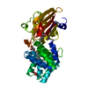
| ||||||||
|---|---|---|---|---|---|---|---|---|---|
| 1 |
| ||||||||
| Unit cell |
| ||||||||
| Components on special symmetry positions |
|
-Components
-Protein , 1 types, 1 molecules 1
| #1: Protein | Mass: 35875.887 Da / Num. of mol.: 1 Source method: isolated from a genetically manipulated source Source: (gene. exp.) |
|---|
-Non-polymers , 5 types, 404 molecules 


| #2: Chemical | ChemComp-45D /  Mass: 564.840 Da / Num. of mol.: 1 / Source method: obtained synthetically / Formula: C40H52O2 / Feature type: SUBJECT OF INVESTIGATION Mass: 564.840 Da / Num. of mol.: 1 / Source method: obtained synthetically / Formula: C40H52O2 / Feature type: SUBJECT OF INVESTIGATION |
|---|---|
| #3: Chemical | ChemComp-GOL /  Mass: 92.094 Da / Num. of mol.: 1 / Source method: obtained synthetically / Formula: C3H8O3 Mass: 92.094 Da / Num. of mol.: 1 / Source method: obtained synthetically / Formula: C3H8O3 |
| #4: Chemical | ChemComp-ACT /  Mass: 59.044 Da / Num. of mol.: 1 / Source method: obtained synthetically / Formula: C2H3O2 Mass: 59.044 Da / Num. of mol.: 1 / Source method: obtained synthetically / Formula: C2H3O2 |
| #5: Chemical | ChemComp-CL /  Mass: 35.453 Da / Num. of mol.: 1 / Source method: obtained synthetically / Formula: Cl / Feature type: SUBJECT OF INVESTIGATION Mass: 35.453 Da / Num. of mol.: 1 / Source method: obtained synthetically / Formula: Cl / Feature type: SUBJECT OF INVESTIGATION |
| #6: Water | ChemComp-HOH / Mass: 18.015 Da / Num. of mol.: 400 / Source method: isolated from a natural source / Formula: H2O |
-Details
| Has ligand of interest | Y |
|---|
-Experimental details
-Experiment
| Experiment | Method: X-RAY DIFFRACTION / Number of used crystals: 1 |
|---|
- Sample preparation
| Crystal | Density Matthews: 2.53 Å3/Da / Density % sol: 51.41 % |
|---|---|
| Crystal grow | Temperature: 293 K / Method: vapor diffusion, sitting drop / pH: 4.5 Details: 100 mM sodium acetate pH 4.5, 10 % poly-ethylene glycol 20,000, 3 % glycerol |
-Data collection
| Diffraction | Mean temperature: 100 K / Serial crystal experiment: N |
|---|---|
| Diffraction source | Source: SYNCHROTRON / Site: Diamond / Beamline: I03 / Wavelength: 0.9795 Å |
| Detector | Type: DECTRIS EIGER2 XE 16M / Detector: PIXEL / Date: Jul 4, 2019 |
| Radiation | Monochromator: DIAMOND BEAMLINE I03 / Protocol: SINGLE WAVELENGTH / Monochromatic (M) / Laue (L): M / Scattering type: x-ray |
| Radiation wavelength | Wavelength: 0.9795 Å / Relative weight: 1 |
| Reflection | Resolution: 1.399→55.542 Å / Num. obs: 68765 / % possible obs: 100 % / Redundancy: 19.8 % / Rmerge(I) obs: 0.133 / Rpim(I) all: 0.044 / Rrim(I) all: 0.14 / Net I/σ(I): 14.4 |
| Reflection shell | Resolution: 1.4→1.44 Å / Rmerge(I) obs: 9.9 / Num. unique obs: 5049 / Rpim(I) all: 3.645 / Rrim(I) all: 11.51 |
- Processing
| Software |
| |||||||||||||||||||||||||||||||||||||||||||||||||||||||||||||||||||||||||||||||||||||||||||||||||||||||||||||||||||||||||||||||||||||||||||||||||||
|---|---|---|---|---|---|---|---|---|---|---|---|---|---|---|---|---|---|---|---|---|---|---|---|---|---|---|---|---|---|---|---|---|---|---|---|---|---|---|---|---|---|---|---|---|---|---|---|---|---|---|---|---|---|---|---|---|---|---|---|---|---|---|---|---|---|---|---|---|---|---|---|---|---|---|---|---|---|---|---|---|---|---|---|---|---|---|---|---|---|---|---|---|---|---|---|---|---|---|---|---|---|---|---|---|---|---|---|---|---|---|---|---|---|---|---|---|---|---|---|---|---|---|---|---|---|---|---|---|---|---|---|---|---|---|---|---|---|---|---|---|---|---|---|---|---|---|---|---|
| Refinement | Method to determine structure: MOLECULAR REPLACEMENT Starting model: 4XB5 Resolution: 1.399→55.542 Å / Cor.coef. Fo:Fc: 0.969 / Cor.coef. Fo:Fc free: 0.963 / WRfactor Rfree: 0.21 / WRfactor Rwork: 0.197 / SU B: 2.08 / SU ML: 0.079 / Average fsc free: 0.9106 / Average fsc work: 0.9181 / Cross valid method: THROUGHOUT / ESU R: 0.1 / ESU R Free: 0.086 / Details: Hydrogens have not been used
| |||||||||||||||||||||||||||||||||||||||||||||||||||||||||||||||||||||||||||||||||||||||||||||||||||||||||||||||||||||||||||||||||||||||||||||||||||
| Solvent computation | Ion probe radii: 0.8 Å / Shrinkage radii: 0.8 Å / VDW probe radii: 1.2 Å / Solvent model: MASK BULK SOLVENT | |||||||||||||||||||||||||||||||||||||||||||||||||||||||||||||||||||||||||||||||||||||||||||||||||||||||||||||||||||||||||||||||||||||||||||||||||||
| Displacement parameters | Biso mean: 25.71 Å2
| |||||||||||||||||||||||||||||||||||||||||||||||||||||||||||||||||||||||||||||||||||||||||||||||||||||||||||||||||||||||||||||||||||||||||||||||||||
| Refinement step | Cycle: LAST / Resolution: 1.399→55.542 Å
| |||||||||||||||||||||||||||||||||||||||||||||||||||||||||||||||||||||||||||||||||||||||||||||||||||||||||||||||||||||||||||||||||||||||||||||||||||
| Refine LS restraints |
| |||||||||||||||||||||||||||||||||||||||||||||||||||||||||||||||||||||||||||||||||||||||||||||||||||||||||||||||||||||||||||||||||||||||||||||||||||
| LS refinement shell |
|
