 ムービー
ムービー コントローラー


+ データを開く

- 基本情報
基本情報
| 登録情報 | データベース: PDB / ID: 7zpf | ||||||
|---|---|---|---|---|---|---|---|
| タイトル | Three-dimensional structure of AIP56, a short-trip single chain AB toxin from Photobacterium damselae subsp. piscicida. | ||||||
 要素 要素 | Aip56 | ||||||
 キーワード キーワード | TOXIN / Apoptosis-inducing protein of 56 kDa / AB-toxin / zinc-metalloprotease / NF-kB p65 target | ||||||
| 機能・相同性 | NFkB-p65-degrading zinc protease / NFkB-p65-degrading zinc protease / metal ion binding / NICKEL (II) ION / Aip56 機能・相同性情報 機能・相同性情報 | ||||||
| 生物種 |  Photobacterium damselae subsp. piscicida (バクテリア) Photobacterium damselae subsp. piscicida (バクテリア) | ||||||
| 手法 |  X線回折 / シンクロトロン / 分子置換 / 解像度: 2.54 Å X線回折 / シンクロトロン / 分子置換 / 解像度: 2.54 Å | ||||||
 データ登録者 データ登録者 | Lisboa, J. / Pereira, P.J.B. / dos Santos, N.M.S. | ||||||
| 資金援助 |  ポルトガル, 1件 ポルトガル, 1件
| ||||||
 引用 引用 | ジャーナル: Nat Commun / 年: 2023 タイトル: Unconventional structure and mechanisms for membrane interaction and translocation of the NF-kappa B-targeting toxin AIP56. 著者: Lisboa, J. / Pereira, C. / Pinto, R.D. / Rodrigues, I.S. / Pereira, L.M.G. / Pinheiro, B. / Oliveira, P. / Pereira, P.J.B. / Azevedo, J.E. / Durand, D. / Benz, R. / do Vale, A. / Dos Santos, N.M.S. | ||||||
| 履歴 |
|
- 構造の表示
構造の表示
| 構造ビューア | 分子: MolmilJmol/JSmol |
|---|
- ダウンロードとリンク
-ダウンロード
| PDBx/mmCIF形式 | 7zpf.cif.gz | 743.8 KB | 表示 | PDBx/mmCIF形式 |
|---|---|---|---|---|
| PDB形式 | pdb7zpf.ent.gz | 620.7 KB | 表示 | PDB形式 |
| PDBx/mmJSON形式 | 7zpf.json.gz | ツリー表示 | PDBx/mmJSON形式 | |
| その他 |  その他のダウンロード その他のダウンロード |
-検証レポート
| 文書・要旨 | 7zpf_validation.pdf.gz | 480.7 KB | 表示 | wwPDB検証レポート |
|---|---|---|---|---|
| 文書・詳細版 | 7zpf_full_validation.pdf.gz | 510.4 KB | 表示 | |
| XML形式データ | 7zpf_validation.xml.gz | 63.8 KB | 表示 | |
| CIF形式データ | 7zpf_validation.cif.gz | 87.4 KB | 表示 | |
| アーカイブディレクトリ | https://data.pdbj.org/pub/pdb/validation_reports/zp/7zpfftp://data.pdbj.org/pub/pdb/validation_reports/zp/7zpf | HTTPS FTP |
-関連構造データ
| 類似構造データ | |
|---|---|
| その他のデータベース |
|
-リンク
 PDBj
PDBj- 集合体
集合体
| 登録構造単位 | 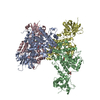
| ||||||||
|---|---|---|---|---|---|---|---|---|---|
| 1 | 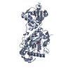
| ||||||||
| 2 | 
| ||||||||
| 3 | 
| ||||||||
| 4 | 
| ||||||||
| 単位格子 |
|
-要素
| #1: タンパク質 | 分子量: 59011.941 Da / 分子数: 4 / 由来タイプ: 組換発現 由来: (組換発現) Photobacterium damselae subsp. piscicida (バクテリア)遺伝子: aip56, E4T25_16935 / 発現宿主: #2: 化合物 | ChemComp-ZN /   分子量: 65.409 Da / 分子数: 4 / 由来タイプ: 合成 / 式: Zn 分子量: 65.409 Da / 分子数: 4 / 由来タイプ: 合成 / 式: Zn#3: 化合物 | ChemComp-NI /   分子量: 58.693 Da / 分子数: 4 / 由来タイプ: 合成 / 式: Ni 分子量: 58.693 Da / 分子数: 4 / 由来タイプ: 合成 / 式: Ni#4: 化合物 | ChemComp-GOL /   分子量: 92.094 Da / 分子数: 5 / 由来タイプ: 合成 / 式: C3H8O3 分子量: 92.094 Da / 分子数: 5 / 由来タイプ: 合成 / 式: C3H8O3#5: 水 | ChemComp-HOH / |  分子量: 18.015 Da / 分子数: 207 / 由来タイプ: 天然 / 式: H2O 分子量: 18.015 Da / 分子数: 207 / 由来タイプ: 天然 / 式: H2O研究の焦点であるリガンドがあるか | N | Has protein modification | Y | |
|---|
-実験情報
-実験
| 実験 | 手法: X線回折 / 使用した結晶の数: 1 |
|---|
- 試料調製
試料調製
| 結晶 | マシュー密度: 2.57 Å3/Da / 溶媒含有率: 52.24 % |
|---|---|
| 結晶化 | 温度: 293 K / 手法: 蒸気拡散法, ハンギングドロップ法 / pH: 8.5 詳細: 400 mM sodium acetate, 100 mM Tris pH 8.5, 15 % PEG 4K, 10 mM Taurine |
-データ収集
| 回折 | 平均測定温度: 100 K / Serial crystal experiment: N |
|---|---|
| 放射光源 | 由来: シンクロトロン / サイト: ESRF  / ビームライン: ID29 / 波長: 0.972 Å / ビームライン: ID29 / 波長: 0.972 Å |
| 検出器 | タイプ: DECTRIS PILATUS 6M / 検出器: PIXEL / 日付: 2015年5月10日 |
| 放射 | プロトコル: SINGLE WAVELENGTH / 単色(M)・ラウエ(L): M / 散乱光タイプ: x-ray |
| 放射波長 | 波長: 0.972 Å / 相対比: 1 |
| 反射 | 解像度: 2.54→46.28 Å / Num. obs: 52848 / % possible obs: 93.4 % / 冗長度: 5 % / Biso Wilson estimate: 48.9 Å2 / CC1/2: 0.992 / Rmerge(I) obs: 0.163 / Rpim(I) all: 0.081 / Rrim(I) all: 0.182 / Net I/σ(I): 6.7 |
| 反射 シェル | 解像度: 2.54→2.78 Å / Rmerge(I) obs: 1.036 / Num. unique obs: 2640 / CC1/2: 0.488 / Rpim(I) all: 0.509 / Rrim(I) all: 1.158 |
- 解析
解析
| ソフトウェア |
| |||||||||||||||||||||||||||||||||||||||||||||||||||||||||||||||||||||||||||||||||||||||||||||||||||||||||||||||||||||||||||||
|---|---|---|---|---|---|---|---|---|---|---|---|---|---|---|---|---|---|---|---|---|---|---|---|---|---|---|---|---|---|---|---|---|---|---|---|---|---|---|---|---|---|---|---|---|---|---|---|---|---|---|---|---|---|---|---|---|---|---|---|---|---|---|---|---|---|---|---|---|---|---|---|---|---|---|---|---|---|---|---|---|---|---|---|---|---|---|---|---|---|---|---|---|---|---|---|---|---|---|---|---|---|---|---|---|---|---|---|---|---|---|---|---|---|---|---|---|---|---|---|---|---|---|---|---|---|---|
| 精密化 | 構造決定の手法: 分子置換 開始モデル: AlphaFold2 解像度: 2.54→46.28 Å / Cor.coef. Fo:Fc: 0.895 / Cor.coef. Fo:Fc free: 0.859 / 交差検証法: THROUGHOUT / SU Rfree Blow DPI: 0.411
| |||||||||||||||||||||||||||||||||||||||||||||||||||||||||||||||||||||||||||||||||||||||||||||||||||||||||||||||||||||||||||||
| 原子変位パラメータ | Biso mean: 65.95 Å2
| |||||||||||||||||||||||||||||||||||||||||||||||||||||||||||||||||||||||||||||||||||||||||||||||||||||||||||||||||||||||||||||
| Refine analyze | Luzzati coordinate error obs: 0.42 Å | |||||||||||||||||||||||||||||||||||||||||||||||||||||||||||||||||||||||||||||||||||||||||||||||||||||||||||||||||||||||||||||
| 精密化ステップ | サイクル: LAST / 解像度: 2.54→46.28 Å
| |||||||||||||||||||||||||||||||||||||||||||||||||||||||||||||||||||||||||||||||||||||||||||||||||||||||||||||||||||||||||||||
| 拘束条件 |
| |||||||||||||||||||||||||||||||||||||||||||||||||||||||||||||||||||||||||||||||||||||||||||||||||||||||||||||||||||||||||||||
| LS精密化 シェル | 解像度: 2.54→2.71 Å
| |||||||||||||||||||||||||||||||||||||||||||||||||||||||||||||||||||||||||||||||||||||||||||||||||||||||||||||||||||||||||||||
| 精密化 TLS | Refine-ID: X-RAY DIFFRACTION
| |||||||||||||||||||||||||||||||||||||||||||||||||||||||||||||||||||||||||||||||||||||||||||||||||||||||||||||||||||||||||||||
| 精密化 TLSグループ |
|
