 Movie
Movie Controller

[English] 日本語
 Yorodumi
- PDB-7zmc: Ketosynthase domain of module 4 from Brevibacillus Brevis orphan BGC11 -
+ Open data

- Basic information
| Entry | Database: PDB / ID: 7zmc | ||||||
|---|---|---|---|---|---|---|---|
| Title | Ketosynthase domain of module 4 from Brevibacillus Brevis orphan BGC11 | ||||||
 Components Components | Putative polyketide synthase | ||||||
 Keywords Keywords | BIOSYNTHETIC PROTEIN / Ketosynthase / polyketide synthase / thiolase fold / Claisen Condensation | ||||||
| Function / homology |  Function and homology information Function and homology informationDIM/DIP cell wall layer assembly / fatty acid synthase activity / phosphopantetheine binding / 3-oxoacyl-[acyl-carrier-protein] synthase activity / fatty acid biosynthetic process / metal ion binding / plasma membrane / cytoplasm Similarity search - Function | ||||||
| Biological species |  Brevibacillus brevis NBRC 100599 (bacteria) Brevibacillus brevis NBRC 100599 (bacteria) | ||||||
| Method |  X-RAY DIFFRACTION / SYNCHROTRON / MOLECULAR REPLACEMENT / Resolution: 3.1 Å X-RAY DIFFRACTION / SYNCHROTRON / MOLECULAR REPLACEMENT / Resolution: 3.1 Å | ||||||
 Authors Authors | Tittes, Y.U. / Herbst, D.A. / Jakob, R.P. / Maier, T. | ||||||
| Funding support |  Switzerland, 1items Switzerland, 1items
| ||||||
 Citation Citation | Journal: Sci Adv / Year: 2022 Title: The structure of a polyketide synthase bimodule core. Authors: Yves U Tittes / Dominik A Herbst / Solène F X Martin / Hugo Munoz-Hernandez / Roman P Jakob / Timm Maier / Abstract: Polyketide synthases (PKSs) are predominantly microbial biosynthetic enzymes. They assemble highly potent bioactive natural products from simple carboxylic acid precursors. The most versatile ...Polyketide synthases (PKSs) are predominantly microbial biosynthetic enzymes. They assemble highly potent bioactive natural products from simple carboxylic acid precursors. The most versatile families of PKSs are organized as assembly lines of functional modules. Each module performs one round of precursor extension and optional modification, followed by directed transfer of the intermediate to the next module. While enzymatic domains and even modules of PKSs are well understood, the higher-order modular architecture of PKS assembly lines remains elusive. Here, we visualize a PKS bimodule core using cryo-electron microscopy and resolve a two-dimensional meshwork of the bimodule core formed by homotypic interactions between modules. The sheet-like organization provides the framework for efficient substrate transfer and for sequestration of trans-acting enzymes required for polyketide production. | ||||||
| History |
|
- Structure visualization
| Structure viewer | Molecule: MolmilJmol/JSmol |
|---|
- Downloads & links
-Download
| PDBx/mmCIF format | 7zmc.cif.gz | 733.6 KB | Display | PDBx/mmCIF format |
|---|---|---|---|---|
| PDB format | pdb7zmc.ent.gz | 519.1 KB | Display | PDB format |
| PDBx/mmJSON format | 7zmc.json.gz | Tree view | PDBx/mmJSON format | |
| Others |  Other downloads Other downloads |
-Validation report
| Arichive directory | https://data.pdbj.org/pub/pdb/validation_reports/zm/7zmcftp://data.pdbj.org/pub/pdb/validation_reports/zm/7zmc | HTTPS FTP |
|---|
-Related structure data
| Related structure data | 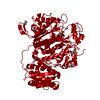 7zm9C  7zmaC  7zmdC  7zmfC  7zskC  4z37S S: Starting model for refinement C: citing same article ( |
|---|---|
| Similar structure data |


-Links
 PDBj
PDBj

- Assembly
| Deposited unit | 
| |||||||||||||||||||||||||||||||||||||||||||||||||||
|---|---|---|---|---|---|---|---|---|---|---|---|---|---|---|---|---|---|---|---|---|---|---|---|---|---|---|---|---|---|---|---|---|---|---|---|---|---|---|---|---|---|---|---|---|---|---|---|---|---|---|---|---|
| 1 |
| |||||||||||||||||||||||||||||||||||||||||||||||||||
| Unit cell |
| |||||||||||||||||||||||||||||||||||||||||||||||||||
| Noncrystallographic symmetry (NCS) | NCS domain:
NCS domain segments:
|
-Components
| #1: Protein | Mass: 70072.117 Da / Num. of mol.: 2 Source method: isolated from a genetically manipulated source Source: (gene. exp.) Brevibacillus brevis NBRC 100599 (bacteria)Strain: 47 / JCM 6285 / NBRC 100599 / Gene: BBR47_39880 / Production host: |
|---|
-Experimental details
-Experiment
| Experiment | Method: X-RAY DIFFRACTION / Number of used crystals: 1 |
|---|
- Sample preparation
| Crystal | Density Matthews: 3.81 Å3/Da / Density % sol: 67.73 % |
|---|---|
| Crystal grow | Temperature: 293 K / Method: vapor diffusion, sitting drop Details: 0.2 ul drops of 4 mg ml-1 protein in buffer (20 mM Hepes KOH pH 8.0, 250 mM NaCl, 5 % v/v glycerol, 5 mM DTT) supplemented by 10 mM MgSO4 with 0.15 ul of reservoir solution (0.2 M (NH4)2SO4, ...Details: 0.2 ul drops of 4 mg ml-1 protein in buffer (20 mM Hepes KOH pH 8.0, 250 mM NaCl, 5 % v/v glycerol, 5 mM DTT) supplemented by 10 mM MgSO4 with 0.15 ul of reservoir solution (0.2 M (NH4)2SO4, 0.1 M Na3 citrate pH 5.22, 8 % w/v PEG 3350) |
-Data collection
| Diffraction | Mean temperature: 100 K / Serial crystal experiment: N |
|---|---|
| Diffraction source | Source: SYNCHROTRON / Site: SLS / Beamline: X06SA / Wavelength: 1 Å |
| Detector | Type: DECTRIS EIGER X 16M / Detector: PIXEL / Date: Oct 5, 2019 |
| Radiation | Protocol: SINGLE WAVELENGTH / Monochromatic (M) / Laue (L): M / Scattering type: x-ray |
| Radiation wavelength | Wavelength: 1 Å / Relative weight: 1 |
| Reflection | Resolution: 3.1→48.97 Å / Num. obs: 38108 / % possible obs: 99.55 % / Redundancy: 13.61 % / Biso Wilson estimate: 105.18 Å2 / CC1/2: 0.999 / Net I/σ(I): 8.44 |
| Reflection shell | Resolution: 3.1→3.28 Å / Redundancy: 0.53 % / Num. unique obs: 3437 / CC1/2: 0.419 / % possible all: 99.8 |
- Processing
| Software |
| |||||||||||||||||||||||||||||||||||||||||||||||||||||||||||||||||||||||||||||||||||||||||||||||||||||||||
|---|---|---|---|---|---|---|---|---|---|---|---|---|---|---|---|---|---|---|---|---|---|---|---|---|---|---|---|---|---|---|---|---|---|---|---|---|---|---|---|---|---|---|---|---|---|---|---|---|---|---|---|---|---|---|---|---|---|---|---|---|---|---|---|---|---|---|---|---|---|---|---|---|---|---|---|---|---|---|---|---|---|---|---|---|---|---|---|---|---|---|---|---|---|---|---|---|---|---|---|---|---|---|---|---|---|---|
| Refinement | Method to determine structure: MOLECULAR REPLACEMENT Starting model: 4z37 Resolution: 3.1→48.97 Å / SU ML: 0.5529 / Cross valid method: FREE R-VALUE / σ(F): 1.33 / Phase error: 37.2566 Stereochemistry target values: GeoStd + Monomer Library + CDL v1.2
| |||||||||||||||||||||||||||||||||||||||||||||||||||||||||||||||||||||||||||||||||||||||||||||||||||||||||
| Solvent computation | Shrinkage radii: 0.9 Å / VDW probe radii: 1.11 Å / Solvent model: FLAT BULK SOLVENT MODEL | |||||||||||||||||||||||||||||||||||||||||||||||||||||||||||||||||||||||||||||||||||||||||||||||||||||||||
| Displacement parameters | Biso mean: 125.72 Å2 | |||||||||||||||||||||||||||||||||||||||||||||||||||||||||||||||||||||||||||||||||||||||||||||||||||||||||
| Refinement step | Cycle: LAST / Resolution: 3.1→48.97 Å
| |||||||||||||||||||||||||||||||||||||||||||||||||||||||||||||||||||||||||||||||||||||||||||||||||||||||||
| Refine LS restraints |
| |||||||||||||||||||||||||||||||||||||||||||||||||||||||||||||||||||||||||||||||||||||||||||||||||||||||||
| Refine LS restraints NCS | Type: Torsion NCS / Rms dev position: 1.3082431265 Å | |||||||||||||||||||||||||||||||||||||||||||||||||||||||||||||||||||||||||||||||||||||||||||||||||||||||||
| LS refinement shell |
| |||||||||||||||||||||||||||||||||||||||||||||||||||||||||||||||||||||||||||||||||||||||||||||||||||||||||
| Refinement TLS params. | Method: refined / Origin x: -31.2516773599 Å / Origin y: -16.3406511113 Å / Origin z: 37.0372630502 Å
| |||||||||||||||||||||||||||||||||||||||||||||||||||||||||||||||||||||||||||||||||||||||||||||||||||||||||
| Refinement TLS group | Refine-ID: X-RAY DIFFRACTION / Refine TLS-ID: 1 / Selection details: all
|
