 Movie
Movie Controller

[English] 日本語
 Yorodumi
- PDB-7zm3: Crystal structure of HsaD from Mycobacterium tuberculosis in comp... -
+ Open data

- Basic information
| Entry | Database: PDB / ID: 7zm3 | ||||||
|---|---|---|---|---|---|---|---|
| Title | Crystal structure of HsaD from Mycobacterium tuberculosis in complex with Cyclipostin-like inhibitor CyC17 | ||||||
 Components Components | 4,5:9,10-diseco-3-hydroxy-5,9,17-trioxoandrosta-1(10),2-diene-4-oate hydrolase | ||||||
 Keywords Keywords | HYDROLASE / HsaD / M. tuberculosis / cholesterol / inhibitor | ||||||
| Function / homology |  Function and homology information Function and homology information4,5:9,10-diseco-3-hydroxy-5,9,17-trioxoandrosta-1(10),2-diene-4-oate hydrolase / 4,5-9,10-diseco-3-hydroxy-5,9,17-trioxoandrosta-1(10),2-diene-4-oate hydrolase activity / 2,6-dioxo-6-phenylhexa-3-enoate hydrolase activity / 2,6-dioxo-6-phenylhexa-3-enoate hydrolase / hydrolase activity, acting on carbon-carbon bonds, in ketonic substances / steroid biosynthetic process / biological process involved in interaction with host / lipid catabolic process / peptidoglycan-based cell wall / hydrolase activity / plasma membrane Similarity search - Function | ||||||
| Biological species |  Mycobacterium tuberculosis H37Rv (bacteria) Mycobacterium tuberculosis H37Rv (bacteria) | ||||||
| Method |  X-RAY DIFFRACTION / SYNCHROTRON / MOLECULAR REPLACEMENT / Resolution: 1.81 Å X-RAY DIFFRACTION / SYNCHROTRON / MOLECULAR REPLACEMENT / Resolution: 1.81 Å | ||||||
 Authors Authors | Barelier, S. / Roig-Zamboni, V. / Cavalier, J.F. / Sulzenbacher, G. | ||||||
| Funding support |  France, 1items France, 1items
| ||||||
 Citation Citation | Journal: Febs J. / Year: 2023 Title: Direct capture, inhibition and crystal structure of HsaD (Rv3569c) from M. tuberculosis. Authors: Barelier, S. / Avellan, R. / Gnawali, G.R. / Fourquet, P. / Roig-Zamboni, V. / Poncin, I. / Point, V. / Bourne, Y. / Audebert, S. / Camoin, L. / Spilling, C.D. / Canaan, S. / Cavalier, J.F. / Sulzenbacher, G. | ||||||
| History |
|
- Structure visualization
| Structure viewer | Molecule: MolmilJmol/JSmol |
|---|
- Downloads & links
-Download
| PDBx/mmCIF format | 7zm3.cif.gz | 130.7 KB | Display | PDBx/mmCIF format |
|---|---|---|---|---|
| PDB format | pdb7zm3.ent.gz | 99.8 KB | Display | PDB format |
| PDBx/mmJSON format | 7zm3.json.gz | Tree view | PDBx/mmJSON format | |
| Others |  Other downloads Other downloads |
-Validation report
| Arichive directory | https://data.pdbj.org/pub/pdb/validation_reports/zm/7zm3ftp://data.pdbj.org/pub/pdb/validation_reports/zm/7zm3 | HTTPS FTP |
|---|
-Related structure data
| Related structure data |  7zjtSC  7zm1C  7zm2C 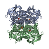 7zm4C S: Starting model for refinement C: citing same article ( |
|---|---|
| Similar structure data |
-Links
 PDBj
PDBj


- Assembly
| Deposited unit | 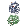
| ||||||||
|---|---|---|---|---|---|---|---|---|---|
| 1 | 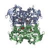
| ||||||||
| Unit cell |
| ||||||||
| Components on special symmetry positions |
|
-Components
| #1: Protein | Mass: 32986.594 Da / Num. of mol.: 2 Source method: isolated from a genetically manipulated source Source: (gene. exp.) Mycobacterium tuberculosis H37Rv (bacteria)Gene: hsaD, bphD, Rv3569c Production host: Mycolicibacterium smegmatis MC2 155 (bacteria)References: UniProt: P9WNH5, 4,5:9,10-diseco-3-hydroxy-5,9,17-trioxoandrosta-1(10),2-diene-4-oate hydrolase, 2,6-dioxo-6-phenylhexa-3-enoate hydrolase #2: Chemical |   Mass: 322.420 Da / Num. of mol.: 2 / Source method: obtained synthetically / Formula: C16H35O4P / Feature type: SUBJECT OF INVESTIGATION Mass: 322.420 Da / Num. of mol.: 2 / Source method: obtained synthetically / Formula: C16H35O4P / Feature type: SUBJECT OF INVESTIGATION#3: Chemical |   Mass: 96.063 Da / Num. of mol.: 2 / Source method: obtained synthetically / Formula: SO4 Mass: 96.063 Da / Num. of mol.: 2 / Source method: obtained synthetically / Formula: SO4#4: Water | ChemComp-HOH / |  Mass: 18.015 Da / Num. of mol.: 336 / Source method: isolated from a natural source / Formula: H2O Mass: 18.015 Da / Num. of mol.: 336 / Source method: isolated from a natural source / Formula: H2OHas ligand of interest | Y | Has protein modification | Y | |
|---|
-Experimental details
-Experiment
| Experiment | Method: X-RAY DIFFRACTION / Number of used crystals: 1 |
|---|
- Sample preparation
| Crystal | Density Matthews: 2.7 Å3/Da / Density % sol: 54.54 % |
|---|---|
| Crystal grow | Temperature: 293 K / Method: vapor diffusion, hanging drop / pH: 6.5 / Details: MES Na 0.1 M NH4SO4 1.656 M PEG400 6.88% |
-Data collection
| Diffraction | Mean temperature: 100 K / Serial crystal experiment: N | ||||||||||||||||||||||||||||||
|---|---|---|---|---|---|---|---|---|---|---|---|---|---|---|---|---|---|---|---|---|---|---|---|---|---|---|---|---|---|---|---|
| Diffraction source | Source: SYNCHROTRON / Site: SOLEIL / Beamline: PROXIMA 2 / Wavelength: 0.980114 Å | ||||||||||||||||||||||||||||||
| Detector | Type: DECTRIS EIGER X 9M / Detector: PIXEL / Date: Jul 27, 2021 | ||||||||||||||||||||||||||||||
| Radiation | Protocol: SINGLE WAVELENGTH / Monochromatic (M) / Laue (L): M / Scattering type: x-ray | ||||||||||||||||||||||||||||||
| Radiation wavelength | Wavelength: 0.980114 Å / Relative weight: 1 | ||||||||||||||||||||||||||||||
| Reflection | Resolution: 1.81→45.36 Å / Num. obs: 66020 / % possible obs: 99.8 % / Redundancy: 13.4 % / Biso Wilson estimate: 19.116 Å2 / CC1/2: 0.999 / Rmerge(I) obs: 0.118 / Rpim(I) all: 0.033 / Rrim(I) all: 0.122 / Net I/σ(I): 16.5 | ||||||||||||||||||||||||||||||
| Reflection shell | Diffraction-ID: 1
|
- Processing
| Software |
| ||||||||||||||||||||||||||||||||||||||||||||||||||||||||||||
|---|---|---|---|---|---|---|---|---|---|---|---|---|---|---|---|---|---|---|---|---|---|---|---|---|---|---|---|---|---|---|---|---|---|---|---|---|---|---|---|---|---|---|---|---|---|---|---|---|---|---|---|---|---|---|---|---|---|---|---|---|---|
| Refinement | Method to determine structure: MOLECULAR REPLACEMENT Starting model: 7ZJT Resolution: 1.81→45.36 Å / Cor.coef. Fo:Fc: 0.962 / Cor.coef. Fo:Fc free: 0.951 / SU B: 2.573 / SU ML: 0.077 / Cross valid method: THROUGHOUT / σ(F): 0 / ESU R: 0.111 / ESU R Free: 0.108 / Stereochemistry target values: MAXIMUM LIKELIHOOD Details: HYDROGENS HAVE BEEN ADDED IN THE RIDING POSITIONS U VALUES : REFINED INDIVIDUALLY
| ||||||||||||||||||||||||||||||||||||||||||||||||||||||||||||
| Solvent computation | Ion probe radii: 0.8 Å / Shrinkage radii: 0.8 Å / VDW probe radii: 1.2 Å / Solvent model: MASK | ||||||||||||||||||||||||||||||||||||||||||||||||||||||||||||
| Displacement parameters | Biso max: 87.71 Å2 / Biso mean: 26.724 Å2 / Biso min: 10.51 Å2
| ||||||||||||||||||||||||||||||||||||||||||||||||||||||||||||
| Refinement step | Cycle: final / Resolution: 1.81→45.36 Å
| ||||||||||||||||||||||||||||||||||||||||||||||||||||||||||||
| Refine LS restraints |
| ||||||||||||||||||||||||||||||||||||||||||||||||||||||||||||
| LS refinement shell | Resolution: 1.81→1.855 Å / Rfactor Rfree error: 0
|
