 Movie
Movie Controller

[English] 日本語
 Yorodumi
- PDB-7zjr: X-ray structure of the periplasmic region of PilJ from P. aeruginosa -
+ Open data

- Basic information
| Entry | Database: PDB / ID: 7zjr | ||||||
|---|---|---|---|---|---|---|---|
| Title | X-ray structure of the periplasmic region of PilJ from P. aeruginosa | ||||||
 Components Components | Protein PilJ | ||||||
 Keywords Keywords | SIGNALING PROTEIN / pilJ / Signal mechanotransduction / chemoreceptor-like / twitching motility protein / two-components system / methyl-accepting-chemotaxis-protein MCP homolog | ||||||
| Function / homology |  Function and homology information Function and homology informationchemotaxis / transmembrane signaling receptor activity / signal transduction / plasma membrane Similarity search - Function | ||||||
| Biological species |  Pseudomonas aeruginosa PAO1 (bacteria) Pseudomonas aeruginosa PAO1 (bacteria) | ||||||
| Method |  X-RAY DIFFRACTION / SYNCHROTRON / MOLECULAR REPLACEMENT / Resolution: 2.50312151016 Å X-RAY DIFFRACTION / SYNCHROTRON / MOLECULAR REPLACEMENT / Resolution: 2.50312151016 Å | ||||||
 Authors Authors | Pierrat, X. / Pojer, F. / Persat, A. | ||||||
| Funding support |  Switzerland, 1items Switzerland, 1items
| ||||||
 Citation Citation | Journal: To Be Published Title: X-ray structure of the periplasmic region of PilJ from P. aeruginosa Authors: Pierrat, X. / Pojer, F. / Persat, A. | ||||||
| History |
|
- Structure visualization
| Structure viewer | Molecule: MolmilJmol/JSmol |
|---|
- Downloads & links
-Download
| PDBx/mmCIF format | 7zjr.cif.gz | 76.4 KB | Display | PDBx/mmCIF format |
|---|---|---|---|---|
| PDB format | pdb7zjr.ent.gz | 45.6 KB | Display | PDB format |
| PDBx/mmJSON format | 7zjr.json.gz | Tree view | PDBx/mmJSON format | |
| Others |  Other downloads Other downloads |
-Validation report
| Summary document | 7zjr_validation.pdf.gz | 422.4 KB | Display | wwPDB validaton report |
|---|---|---|---|---|
| Full document | 7zjr_full_validation.pdf.gz | 424 KB | Display | |
| Data in XML | 7zjr_validation.xml.gz | 10.9 KB | Display | |
| Data in CIF | 7zjr_validation.cif.gz | 14.1 KB | Display | |
| Arichive directory | https://data.pdbj.org/pub/pdb/validation_reports/zj/7zjrftp://data.pdbj.org/pub/pdb/validation_reports/zj/7zjr | HTTPS FTP |
-Related structure data
| Related structure data |  2yfaS S: Starting model for refinement |
|---|---|
| Similar structure data |
-Links
 PDBj
PDBj

- Assembly
| Deposited unit | 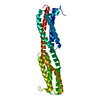
| ||||||||||||
|---|---|---|---|---|---|---|---|---|---|---|---|---|---|
| 1 |
| ||||||||||||
| Unit cell |
|
-Components
| #1: Protein | Mass: 31759.145 Da / Num. of mol.: 1 Source method: isolated from a genetically manipulated source Source: (gene. exp.) Pseudomonas aeruginosa PAO1 (bacteria) / Gene: pilJ, PA0411 / Plasmid: pXP209 - 6xHis_Thrombin_PilJ_periplasmic / Details (production host): PilJ(36-309) / Production host: |
|---|---|
| #2: Water | ChemComp-HOH /  Mass: 18.015 Da / Num. of mol.: 14 / Source method: isolated from a natural source / Formula: H2O Mass: 18.015 Da / Num. of mol.: 14 / Source method: isolated from a natural source / Formula: H2O |
-Experimental details
-Experiment
| Experiment | Method: X-RAY DIFFRACTION / Number of used crystals: 1 |
|---|
- Sample preparation
| Crystal grow | Temperature: 291 K / Method: vapor diffusion, sitting drop / Details: 30% v/v Jeffamine M-600 pH 7.0, 0.1 M Tris pH 8.5 |
|---|
-Data collection
| Diffraction | Mean temperature: 100 K / Serial crystal experiment: N |
|---|---|
| Diffraction source | Source: SYNCHROTRON / Site: SLS / Beamline: X06DA / Wavelength: 1 Å |
| Detector | Type: DECTRIS PILATUS 2M-F / Detector: PIXEL / Date: Sep 4, 2018 |
| Radiation | Protocol: SINGLE WAVELENGTH / Monochromatic (M) / Laue (L): M / Scattering type: x-ray |
| Radiation wavelength | Wavelength: 1 Å / Relative weight: 1 |
| Reflection | Resolution: 2.32→49.4400799314 Å / Num. obs: 54408 / % possible obs: 99.9 % / Observed criterion σ(F): 1 / Redundancy: 7.8 % / Biso Wilson estimate: 64.6291655926 Å2 / Rrim(I) all: 0.068 / Net I/σ(I): 21.36 |
| Reflection shell | Resolution: 2.32→2.46 Å / Mean I/σ(I) obs: 1.07 / Num. unique obs: 8751 / CC1/2: 0.45 / Rrim(I) all: 1.96 |
- Processing
| Software |
| |||||||||||||||||||||||||||||||||||||||||||||||||||||||||||||||
|---|---|---|---|---|---|---|---|---|---|---|---|---|---|---|---|---|---|---|---|---|---|---|---|---|---|---|---|---|---|---|---|---|---|---|---|---|---|---|---|---|---|---|---|---|---|---|---|---|---|---|---|---|---|---|---|---|---|---|---|---|---|---|---|---|
| Refinement | Method to determine structure: MOLECULAR REPLACEMENT Starting model: 2yfa Resolution: 2.50312151016→49.4400799314 Å / SU ML: 0.364286546272 / Cross valid method: FREE R-VALUE / σ(F): 1.3400428594 / Phase error: 27.4485563934 Stereochemistry target values: GeoStd + Monomer Library + CDL v1.2
| |||||||||||||||||||||||||||||||||||||||||||||||||||||||||||||||
| Solvent computation | Shrinkage radii: 0.9 Å / VDW probe radii: 1.11 Å / Solvent model: FLAT BULK SOLVENT MODEL | |||||||||||||||||||||||||||||||||||||||||||||||||||||||||||||||
| Displacement parameters | Biso mean: 67.1813474692 Å2 | |||||||||||||||||||||||||||||||||||||||||||||||||||||||||||||||
| Refinement step | Cycle: LAST / Resolution: 2.50312151016→49.4400799314 Å
| |||||||||||||||||||||||||||||||||||||||||||||||||||||||||||||||
| Refine LS restraints |
| |||||||||||||||||||||||||||||||||||||||||||||||||||||||||||||||
| LS refinement shell |
|
