 Movie
Movie Controller

[English] 日本語
 Yorodumi
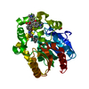
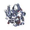
- PDB-7zix: X-ray structure of the haloalkane dehalogenase HaloTag7 bound to ... -
+ Open data

- Basic information
| Entry | Database: PDB / ID: 7zix | ||||||
|---|---|---|---|---|---|---|---|
| Title | X-ray structure of the haloalkane dehalogenase HaloTag7 bound to a butylmethanesulfonamide tetramethylrhodamine ligand (TMR-S4) | ||||||
 Components Components | Haloalkane dehalogenase | ||||||
 Keywords Keywords | HYDROLASE / haloalkane dehalogenase / HaloTag / HaloTag7 / Self-Labeling Protein / Fluorophore / Tetramethylrhodamine | ||||||
| Function / homology |  Function and homology information Function and homology informationhaloalkane dehalogenase / haloalkane dehalogenase activity / response to toxic substance / membrane Similarity search - Function | ||||||
| Biological species |  Rhodococcus sp. (bacteria) Rhodococcus sp. (bacteria) | ||||||
| Method |  X-RAY DIFFRACTION / SYNCHROTRON / MOLECULAR REPLACEMENT / Resolution: 2.39 Å X-RAY DIFFRACTION / SYNCHROTRON / MOLECULAR REPLACEMENT / Resolution: 2.39 Å | ||||||
 Authors Authors | Tarnawski, M. / Kompa, J. / Johnsson, K. / Hiblot, J. | ||||||
| Funding support |  Germany, 1items Germany, 1items
| ||||||
 Citation Citation | Journal: To Be Published Title: X-ray structure of the haloalkane dehalogenase HaloTag7 bound to a butylmethanesulfonamide tetramethylrhodamine ligand (HSAm(4)-TMR) Authors: Tarnawski, M. / Kompa, J. / Johnsson, K. / Hiblot, J. | ||||||
| History |
|
- Structure visualization
| Structure viewer | Molecule: MolmilJmol/JSmol |
|---|
- Downloads & links
-Download
| PDBx/mmCIF format | 7zix.cif.gz | 153.4 KB | Display | PDBx/mmCIF format |
|---|---|---|---|---|
| PDB format | pdb7zix.ent.gz | 102.7 KB | Display | PDB format |
| PDBx/mmJSON format | 7zix.json.gz | Tree view | PDBx/mmJSON format | |
| Others |  Other downloads Other downloads |
-Validation report
| Arichive directory | https://data.pdbj.org/pub/pdb/validation_reports/zi/7zixftp://data.pdbj.org/pub/pdb/validation_reports/zi/7zix | HTTPS FTP |
|---|
-Related structure data
| Related structure data |  6y7aS S: Starting model for refinement |
|---|---|
| Similar structure data |
-Links
 PDBj
PDBj


- Assembly
| Deposited unit | 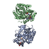
| ||||||||||||
|---|---|---|---|---|---|---|---|---|---|---|---|---|---|
| 1 | 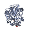
| ||||||||||||
| 2 | 
| ||||||||||||
| Unit cell |
|
-Components
| #1: Protein | Mass: 33225.980 Da / Num. of mol.: 2 Source method: isolated from a genetically manipulated source Source: (gene. exp.) Rhodococcus sp. (bacteria) / Gene: dhaA / Production host: #2: Chemical | 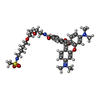  Mass: 667.792 Da / Num. of mol.: 2 / Source method: obtained synthetically / Formula: C34H43N4O8S / Feature type: SUBJECT OF INVESTIGATION Mass: 667.792 Da / Num. of mol.: 2 / Source method: obtained synthetically / Formula: C34H43N4O8S / Feature type: SUBJECT OF INVESTIGATION#3: Water | ChemComp-HOH / |  Mass: 18.015 Da / Num. of mol.: 145 / Source method: isolated from a natural source / Formula: H2O Mass: 18.015 Da / Num. of mol.: 145 / Source method: isolated from a natural source / Formula: H2OHas ligand of interest | Y | |
|---|
-Experimental details
-Experiment
| Experiment | Method: X-RAY DIFFRACTION / Number of used crystals: 1 |
|---|
- Sample preparation
| Crystal | Density Matthews: 2.13 Å3/Da / Density % sol: 42.18 % |
|---|---|
| Crystal grow | Temperature: 293 K / Method: vapor diffusion, hanging drop Details: 0.1 M MES pH 6.0, 1.0 M lithium chloride, 23% (m/v) PEG 6000 |
-Data collection
| Diffraction | Mean temperature: 100 K / Serial crystal experiment: N |
|---|---|
| Diffraction source | Source: SYNCHROTRON / Site: SLS  / Beamline: X10SA / Wavelength: 1.00007 Å / Beamline: X10SA / Wavelength: 1.00007 Å |
| Detector | Type: DECTRIS EIGER2 X 16M / Detector: PIXEL / Date: Dec 16, 2020 |
| Radiation | Monochromator: Si(111) / Protocol: SINGLE WAVELENGTH / Monochromatic (M) / Laue (L): M / Scattering type: x-ray |
| Radiation wavelength | Wavelength: 1.00007 Å / Relative weight: 1 |
| Reflection | Resolution: 2.39→50 Å / Num. obs: 20776 / % possible obs: 93.5 % / Redundancy: 3.83 % / Biso Wilson estimate: 15.93 Å2 / CC1/2: 0.994 / Rmerge(I) obs: 0.123 / Net I/σ(I): 7.5 |
| Reflection shell | Resolution: 2.39→2.45 Å / Redundancy: 3.69 % / Rmerge(I) obs: 0.483 / Mean I/σ(I) obs: 2.7 / Num. unique obs: 1039 / CC1/2: 0.874 / % possible all: 68.5 |
- Processing
| Software |
| |||||||||||||||||||||||||||||||||||||||||||||||||||||||||||||||
|---|---|---|---|---|---|---|---|---|---|---|---|---|---|---|---|---|---|---|---|---|---|---|---|---|---|---|---|---|---|---|---|---|---|---|---|---|---|---|---|---|---|---|---|---|---|---|---|---|---|---|---|---|---|---|---|---|---|---|---|---|---|---|---|---|
| Refinement | Method to determine structure: MOLECULAR REPLACEMENT Starting model: 6Y7A Resolution: 2.39→44.18 Å / SU ML: 0.2158 / Cross valid method: FREE R-VALUE / σ(F): 1.35 / Phase error: 24.5197 Stereochemistry target values: GeoStd + Monomer Library + CDL v1.2
| |||||||||||||||||||||||||||||||||||||||||||||||||||||||||||||||
| Solvent computation | Shrinkage radii: 0.9 Å / VDW probe radii: 1.11 Å / Solvent model: FLAT BULK SOLVENT MODEL | |||||||||||||||||||||||||||||||||||||||||||||||||||||||||||||||
| Displacement parameters | Biso mean: 36.02 Å2 | |||||||||||||||||||||||||||||||||||||||||||||||||||||||||||||||
| Refinement step | Cycle: LAST / Resolution: 2.39→44.18 Å
| |||||||||||||||||||||||||||||||||||||||||||||||||||||||||||||||
| Refine LS restraints |
| |||||||||||||||||||||||||||||||||||||||||||||||||||||||||||||||
| LS refinement shell |
|
