 Movie
Movie Controller


+ Open data

- Basic information
| Entry | Database: PDB / ID: 7zgi | ||||||
|---|---|---|---|---|---|---|---|
| Title | chloroplast trigger factor (TIG1) | ||||||
 Components Components | Peptidylprolyl isomerase | ||||||
 Keywords Keywords | CHAPERONE / Molecular chaperones / chloroplast / co-translational folding / PPIase | ||||||
| Function / homology |  Function and homology information Function and homology information'de novo' cotranslational protein folding / protein unfolding / : / protein folding chaperone / peptidylprolyl isomerase / peptidyl-prolyl cis-trans isomerase activity / protein transport / ribosome binding Similarity search - Function | ||||||
| Biological species |   Chlamydomonas reinhardtii (plant) Chlamydomonas reinhardtii (plant) | ||||||
| Method |  X-RAY DIFFRACTION / SYNCHROTRON / SAD / Resolution: 2.6 Å X-RAY DIFFRACTION / SYNCHROTRON / SAD / Resolution: 2.6 Å | ||||||
 Authors Authors | Carius, Y. / Ries, F. / Gries, K. / Trentmann, O. / Willmund, F. / Lancaster, C.R.D. | ||||||
| Funding support |  Germany, 1items Germany, 1items
| ||||||
 Citation Citation | Journal: Acta Crystallogr D Struct Biol / Year: 2022 Title: Structural features of chloroplast trigger factor determined at 2.6 angstrom resolution. Authors: Carius, Y. / Ries, F. / Gries, K. / Trentmann, O. / Lancaster, C.R.D. / Willmund, F. | ||||||
| History |
|
- Structure visualization
| Structure viewer | Molecule: MolmilJmol/JSmol |
|---|
- Downloads & links
-Download
| PDBx/mmCIF format | 7zgi.cif.gz | 148.4 KB | Display | PDBx/mmCIF format |
|---|---|---|---|---|
| PDB format | pdb7zgi.ent.gz | 115.6 KB | Display | PDB format |
| PDBx/mmJSON format | 7zgi.json.gz | Tree view | PDBx/mmJSON format | |
| Others |  Other downloads Other downloads |
-Validation report
| Summary document | 7zgi_validation.pdf.gz | 443.8 KB | Display | wwPDB validaton report |
|---|---|---|---|---|
| Full document | 7zgi_full_validation.pdf.gz | 453.5 KB | Display | |
| Data in XML | 7zgi_validation.xml.gz | 15.7 KB | Display | |
| Data in CIF | 7zgi_validation.cif.gz | 22.7 KB | Display | |
| Arichive directory | https://data.pdbj.org/pub/pdb/validation_reports/zg/7zgiftp://data.pdbj.org/pub/pdb/validation_reports/zg/7zgi | HTTPS FTP |
-Related structure data
| Similar structure data |
|---|
-Links
 PDBj
PDBj


- Assembly
| Deposited unit | 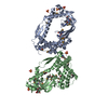
| ||||||||
|---|---|---|---|---|---|---|---|---|---|
| 1 | 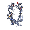
| ||||||||
| 2 | 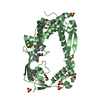
| ||||||||
| Unit cell |
| ||||||||
| Components on special symmetry positions |
|
-Components
| #1: Protein | Mass: 54550.516 Da / Num. of mol.: 2 Source method: isolated from a genetically manipulated source Source: (gene. exp.) Chlamydomonas reinhardtii (plant) / Gene: TIG1, CHLRE_01g001750v5, CHLREDRAFT_196868 / Production host:  #2: Chemical | ChemComp-SO4 /   Mass: 96.063 Da / Num. of mol.: 8 / Source method: obtained synthetically / Formula: SO4 Mass: 96.063 Da / Num. of mol.: 8 / Source method: obtained synthetically / Formula: SO4#3: Chemical | ChemComp-PEG /   Mass: 106.120 Da / Num. of mol.: 4 / Source method: obtained synthetically / Formula: C4H10O3 Mass: 106.120 Da / Num. of mol.: 4 / Source method: obtained synthetically / Formula: C4H10O3#4: Water | ChemComp-HOH / |  Mass: 18.015 Da / Num. of mol.: 26 / Source method: isolated from a natural source / Formula: H2O Mass: 18.015 Da / Num. of mol.: 26 / Source method: isolated from a natural source / Formula: H2OHas ligand of interest | N | Has protein modification | Y | |
|---|
-Experimental details
-Experiment
| Experiment | Method: X-RAY DIFFRACTION / Number of used crystals: 1 |
|---|
- Sample preparation
| Crystal | Density Matthews: 2.82 Å3/Da / Density % sol: 56.35 % |
|---|---|
| Crystal grow | Temperature: 291 K / Method: vapor diffusion, sitting drop / Details: 0.3 M MgSO4, 10 % PEG 3350 |
-Data collection
| Diffraction | Mean temperature: 100 K / Serial crystal experiment: N |
|---|---|
| Diffraction source | Source: SYNCHROTRON / Site: ESRF  / Beamline: ID23-1 / Wavelength: 0.97779 Å / Beamline: ID23-1 / Wavelength: 0.97779 Å |
| Detector | Type: DECTRIS PILATUS 6M-F / Detector: PIXEL / Date: Jan 29, 2017 |
| Radiation | Protocol: SINGLE WAVELENGTH / Monochromatic (M) / Laue (L): M / Scattering type: x-ray |
| Radiation wavelength | Wavelength: 0.97779 Å / Relative weight: 1 |
| Reflection | Resolution: 2.6→54.31 Å / Num. obs: 37064 / % possible obs: 99.8 % / Redundancy: 6.6 % / CC1/2: 0.996 / Rmerge(I) obs: 0.094 / Net I/σ(I): 10.2 |
| Reflection shell | Resolution: 2.6→2.7 Å / Rmerge(I) obs: 0.548 / Num. unique obs: 4515 / CC1/2: 0.941 |
- Processing
| Software |
| ||||||||||||||||||||||||
|---|---|---|---|---|---|---|---|---|---|---|---|---|---|---|---|---|---|---|---|---|---|---|---|---|---|
| Refinement | Method to determine structure: SAD / Resolution: 2.6→54.31 Å / Cor.coef. Fo:Fc: 0.935 / Cor.coef. Fo:Fc free: 0.912 / SU B: 0.004 / SU ML: 0 / Cross valid method: THROUGHOUT / σ(F): 0 / ESU R: 0.241 / ESU R Free: 0.29 / Stereochemistry target values: MAXIMUM LIKELIHOOD Details: HYDROGENS HAVE BEEN ADDED IN THE RIDING POSITIONS U VALUES : REFINED INDIVIDUALLY
| ||||||||||||||||||||||||
| Solvent computation | Ion probe radii: 0.8 Å / Shrinkage radii: 0.8 Å / VDW probe radii: 1.2 Å / Solvent model: MASK | ||||||||||||||||||||||||
| Displacement parameters | Biso max: 146.53 Å2 / Biso mean: 57.871 Å2 / Biso min: 20.37 Å2
| ||||||||||||||||||||||||
| Refinement step | Cycle: final / Resolution: 2.6→54.31 Å
| ||||||||||||||||||||||||
| LS refinement shell | Resolution: 2.6→2.668 Å / Rfactor Rfree error: 0 / Total num. of bins used: 20
|
