 Movie
Movie Controller


+ Open data

- Basic information
| Entry | Database: PDB / ID: 7zet | ||||||
|---|---|---|---|---|---|---|---|
| Title | Crystal structure of human Clusterin, crystal form I | ||||||
 Components Components | Clusterin | ||||||
 Keywords Keywords | CHAPERONE / molecular chaperone / complement system / Alzheimer's disease | ||||||
| Function / homology |  Function and homology information Function and homology informationimmune complex clearance / perinuclear endoplasmic reticulum lumen / regulation of neuronal signal transduction / positive regulation of neurofibrillary tangle assembly / central nervous system myelin maintenance / response to misfolded protein / negative regulation of response to endoplasmic reticulum stress / spherical high-density lipoprotein particle / Terminal pathway of complement / protein targeting to lysosome involved in chaperone-mediated autophagy ...immune complex clearance / perinuclear endoplasmic reticulum lumen / regulation of neuronal signal transduction / positive regulation of neurofibrillary tangle assembly / central nervous system myelin maintenance / response to misfolded protein / negative regulation of response to endoplasmic reticulum stress / spherical high-density lipoprotein particle / Terminal pathway of complement / protein targeting to lysosome involved in chaperone-mediated autophagy / protein carrier chaperone / chromaffin granule / regulation of amyloid-beta clearance / microglial cell proliferation / reverse cholesterol transport / misfolded protein binding / negative regulation of intrinsic apoptotic signaling pathway in response to DNA damage / apical dendrite / protein import / complement activation / positive regulation of ubiquitin-dependent protein catabolic process / negative regulation of amyloid fibril formation / Antimicrobial peptides / neurofibrillary tangle / low-density lipoprotein particle receptor binding / positive regulation of amyloid fibril formation / positive regulation of amyloid-beta formation / negative regulation of release of cytochrome c from mitochondria / amyloid-beta clearance / negative regulation of amyloid-beta formation / complement activation, classical pathway / chaperone-mediated protein complex assembly / negative regulation of protein-containing complex assembly / positive regulation of intrinsic apoptotic signaling pathway / : / intrinsic apoptotic signaling pathway / platelet alpha granule lumen / Regulation of Complement cascade / release of cytochrome c from mitochondria / positive regulation of protein-containing complex assembly / microglial cell activation / positive regulation of NF-kappaB transcription factor activity / positive regulation of receptor-mediated endocytosis / lipid metabolic process / response to virus / tau protein binding / cell morphogenesis / positive regulation of nitric oxide biosynthetic process / positive regulation of tumor necrosis factor production / unfolded protein binding / positive regulation of proteasomal ubiquitin-dependent protein catabolic process / Platelet degranulation / regulation of cell population proliferation / protein-folding chaperone binding / : / amyloid-beta binding / regulation of apoptotic process / blood microparticle / negative regulation of neuron apoptotic process / mitochondrial inner membrane / protein stabilization / positive regulation of apoptotic process / receptor ligand activity / signaling receptor binding / innate immune response / intracellular membrane-bounded organelle / synapse / ubiquitin protein ligase binding / positive regulation of gene expression / protein-containing complex binding / perinuclear region of cytoplasm / cell surface / Golgi apparatus / protein-containing complex / mitochondrion / extracellular space / extracellular exosome / extracellular region / nucleus / cytoplasm / cytosol Similarity search - Function | ||||||
| Biological species |  Homo sapiens (human) Homo sapiens (human) | ||||||
| Method |  X-RAY DIFFRACTION / SYNCHROTRON / MOLECULAR REPLACEMENT / molecular replacement / Resolution: 2.8 Å X-RAY DIFFRACTION / SYNCHROTRON / MOLECULAR REPLACEMENT / molecular replacement / Resolution: 2.8 Å | ||||||
 Authors Authors | Yuste-Checa, P. / Bracher, A. / Hartl, F.U. | ||||||
| Funding support | 1items
| ||||||
 Citation Citation | Journal: Nat.Struct.Mol.Biol. / Year: 2025 Title: Structural analyses define the molecular basis of clusterin chaperone function. Authors: Yuste-Checa, P. / Carvajal, A.I. / Mi, C. / Paatz, S. / Hartl, F.U. / Bracher, A. #1: Journal: Biorxiv / Year: 2024Title: Hydrophobic tails enable diverse functions of the extracellular chaperone clusterin Authors: Yuste-Checa, P. / Carvajal, A.I. / Mi, C. / Paatz, S. / Ulrich Hartl, F. / Bracher, A. | ||||||
| History |
|
- Structure visualization
| Structure viewer | Molecule: MolmilJmol/JSmol |
|---|
- Downloads & links
-Download
| PDBx/mmCIF format | 7zet.cif.gz | 95.6 KB | Display | PDBx/mmCIF format |
|---|---|---|---|---|
| PDB format | pdb7zet.ent.gz | 69.8 KB | Display | PDB format |
| PDBx/mmJSON format | 7zet.json.gz | Tree view | PDBx/mmJSON format | |
| Others |  Other downloads Other downloads |
-Validation report
| Summary document | 7zet_validation.pdf.gz | 947.9 KB | Display | wwPDB validaton report |
|---|---|---|---|---|
| Full document | 7zet_full_validation.pdf.gz | 949.7 KB | Display | |
| Data in XML | 7zet_validation.xml.gz | 15 KB | Display | |
| Data in CIF | 7zet_validation.cif.gz | 19.4 KB | Display | |
| Arichive directory | https://data.pdbj.org/pub/pdb/validation_reports/ze/7zetftp://data.pdbj.org/pub/pdb/validation_reports/ze/7zet | HTTPS FTP |
-Related structure data

-Links
 PDBj
PDBj


- Assembly
| Deposited unit | 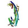
| ||||||||
|---|---|---|---|---|---|---|---|---|---|
| 1 |
| ||||||||
| 2 |
| ||||||||
| Unit cell |
|
-Components
| #1: Protein | Mass: 47038.852 Da / Num. of mol.: 1 / Fragment: Clu-delta(214-238) / Mutation: deletion(214-238) Source method: isolated from a genetically manipulated source Source: (gene. exp.) Homo sapiens (human) / Gene: CLU, APOJ, CLI, KUB1, AAG4 / Plasmid: pB-T-PAF / Cell line (production host): HEK293T / Production host: Homo sapiens (human) / References: UniProt: P10909 | ||||
|---|---|---|---|---|---|
| #2: Polysaccharide | 2-acetamido-2-deoxy-beta-D-glucopyranose-(1-4)-2-acetamido-2-deoxy-beta-D-glucopyranose Source method: isolated from a genetically manipulated source | ||||
| #3: Polysaccharide | beta-D-mannopyranose-(1-4)-2-acetamido-2-deoxy-beta-D-glucopyranose-(1-4)-2-acetamido-2-deoxy-beta- ...beta-D-mannopyranose-(1-4)-2-acetamido-2-deoxy-beta-D-glucopyranose-(1-4)-2-acetamido-2-deoxy-beta-D-glucopyranose Source method: isolated from a genetically manipulated source | ||||
| #4: Sugar | ChemComp-NAG /   Type: D-saccharide, beta linking / Mass: 221.208 Da / Num. of mol.: 4 / Source method: obtained synthetically / Formula: C8H15NO6 Type: D-saccharide, beta linking / Mass: 221.208 Da / Num. of mol.: 4 / Source method: obtained synthetically / Formula: C8H15NO6Has ligand of interest | N | Has protein modification | Y | |
-Experimental details
-Experiment
| Experiment | Method: X-RAY DIFFRACTION / Number of used crystals: 1 |
|---|
- Sample preparation
| Crystal | Density Matthews: 3 Å3/Da / Density % sol: 59.07 % / Mosaicity: 0.25 ° |
|---|---|
| Crystal grow | Temperature: 277 K / Method: vapor diffusion, sitting drop / pH: 5.6 Details: 30% PEG-4000, 0.2 M NH4-acetate, 0.1 M Na-citrate pH 5.6 |
-Data collection
| Diffraction | Mean temperature: 100 K / Serial crystal experiment: N | ||||||||||||||||||||||||||||||
|---|---|---|---|---|---|---|---|---|---|---|---|---|---|---|---|---|---|---|---|---|---|---|---|---|---|---|---|---|---|---|---|
| Diffraction source | Source: SYNCHROTRON / Site: ESRF  / Beamline: ID23-1 / Wavelength: 0.7749 Å / Beamline: ID23-1 / Wavelength: 0.7749 Å | ||||||||||||||||||||||||||||||
| Detector | Type: DECTRIS EIGER X 16M / Detector: PIXEL / Date: Dec 2, 2021 | ||||||||||||||||||||||||||||||
| Radiation | Protocol: SINGLE WAVELENGTH / Monochromatic (M) / Laue (L): M / Scattering type: x-ray | ||||||||||||||||||||||||||||||
| Radiation wavelength | Wavelength: 0.7749 Å / Relative weight: 1 | ||||||||||||||||||||||||||||||
| Reflection | Resolution: 2.8→49.08 Å / Num. obs: 13861 / % possible obs: 98.2 % / Redundancy: 3.7 % / Biso Wilson estimate: 75.02 Å2 / CC1/2: 0.995 / Rmerge(I) obs: 0.146 / Rpim(I) all: 0.088 / Rrim(I) all: 0.172 / Net I/σ(I): 5.9 / Num. measured all: 50668 / Scaling rejects: 4 | ||||||||||||||||||||||||||||||
| Reflection shell | Diffraction-ID: 1
|
-Phasing
| Phasing | Method: molecular replacement | ||||||
|---|---|---|---|---|---|---|---|
| Phasing MR | R rigid body: 0.596
|
- Processing
| Software |
| ||||||||||||||||||||||||||||||||||||||||||
|---|---|---|---|---|---|---|---|---|---|---|---|---|---|---|---|---|---|---|---|---|---|---|---|---|---|---|---|---|---|---|---|---|---|---|---|---|---|---|---|---|---|---|---|
| Refinement | Method to determine structure: MOLECULAR REPLACEMENT Starting model: AlphaFold AF-P10909-F1-model_v1.pdb Resolution: 2.8→27.61 Å / SU ML: 0.54 / Cross valid method: THROUGHOUT / σ(F): 1.34 / Phase error: 35.41 / Stereochemistry target values: ML Details: The Clusterin construct is a glyco-protein, which was expressed in the presence of the alpha-mannosidase I inhibitor kifunensine, which prevents trimming of mannose moieties, resulting in ...Details: The Clusterin construct is a glyco-protein, which was expressed in the presence of the alpha-mannosidase I inhibitor kifunensine, which prevents trimming of mannose moieties, resulting in oligo-mannose N-glycans, and thereby prevents processing to mature complex N-glycans (Chang et al., Structure 15, 267(2007), Fig. 1B). Asn residues 86, 103, 145, 291, 354 and 374 are expected to carry predominantly NAG-NAG-BMA-(3->1)MAN-MAN-MAN (6->1)-MAN (3->1)MAN-MAN (6->1)MAN-MAN N-glycan trees (Chang et al., Structure 15, 267(2007), Fig. 1B).
| ||||||||||||||||||||||||||||||||||||||||||
| Solvent computation | Shrinkage radii: 0.9 Å / VDW probe radii: 1.11 Å / Solvent model: FLAT BULK SOLVENT MODEL | ||||||||||||||||||||||||||||||||||||||||||
| Displacement parameters | Biso max: 161.13 Å2 / Biso mean: 80.9892 Å2 / Biso min: 36.33 Å2 | ||||||||||||||||||||||||||||||||||||||||||
| Refinement step | Cycle: final / Resolution: 2.8→27.61 Å
| ||||||||||||||||||||||||||||||||||||||||||
| LS refinement shell | Refine-ID: X-RAY DIFFRACTION / Rfactor Rfree error: 0 / Total num. of bins used: 5
|
