 Movie
Movie Controller


+ Open data

- Basic information
| Entry | Database: PDB / ID: 7zdn | ||||||
|---|---|---|---|---|---|---|---|
| Title | Human Cyclophilin D in complex with fragment | ||||||
 Components Components | Peptidyl-prolyl cis-trans isomerase F, mitochondrial | ||||||
 Keywords Keywords | ISOMERASE / Cyclophilin D PPIase | ||||||
| Function / homology |  Function and homology information Function and homology informationmitochondrial outer membrane permeabilization involved in programmed cell death / regulation of mitochondrial membrane permeability involved in programmed necrotic cell death / mitochondrial permeability transition pore complex / cellular response to arsenic-containing substance / negative regulation of oxidative phosphorylation / regulation of mitochondrial membrane permeability / cyclosporin A binding / negative regulation of release of cytochrome c from mitochondria / negative regulation of intrinsic apoptotic signaling pathway / cellular response to calcium ion ...mitochondrial outer membrane permeabilization involved in programmed cell death / regulation of mitochondrial membrane permeability involved in programmed necrotic cell death / mitochondrial permeability transition pore complex / cellular response to arsenic-containing substance / negative regulation of oxidative phosphorylation / regulation of mitochondrial membrane permeability / cyclosporin A binding / negative regulation of release of cytochrome c from mitochondria / negative regulation of intrinsic apoptotic signaling pathway / cellular response to calcium ion / response to ischemia / enzyme inhibitor activity / peptidylprolyl isomerase / peptidyl-prolyl cis-trans isomerase activity / cellular response to hydrogen peroxide / protein folding / mitochondrial matrix / apoptotic process / negative regulation of apoptotic process / mitochondrion / membrane / cytoplasm Similarity search - Function | ||||||
| Biological species |  Homo sapiens (human) Homo sapiens (human) | ||||||
| Method |  X-RAY DIFFRACTION / SYNCHROTRON / MOLECULAR REPLACEMENT / Resolution: 1.55 Å X-RAY DIFFRACTION / SYNCHROTRON / MOLECULAR REPLACEMENT / Resolution: 1.55 Å | ||||||
 Authors Authors | Silva, D.O. / Graedler, U. / Bandeiras, T.M. | ||||||
| Funding support | 1items
| ||||||
 Citation Citation | Journal: Int.J.Biol.Macromol. / Year: 2026 Title: Structure-based design of pyrazole derivatives targeting the human Cyclophilin D binding site Authors: Silva, D.O. / Freitas, M.C. / Malta, C.F. / Martins, M.T. / Sousa, P.M. / Matias, P.M. / Schwarz, D. / Ventura, M.R. / Gradler, U. / Bandeiras, T.M. | ||||||
| History |
|
- Structure visualization
| Structure viewer | Molecule: MolmilJmol/JSmol |
|---|
- Downloads & links
-Download
| PDBx/mmCIF format | 7zdn.cif.gz | 168.2 KB | Display | PDBx/mmCIF format |
|---|---|---|---|---|
| PDB format | pdb7zdn.ent.gz | 108.7 KB | Display | PDB format |
| PDBx/mmJSON format | 7zdn.json.gz | Tree view | PDBx/mmJSON format | |
| Others |  Other downloads Other downloads |
-Validation report
| Arichive directory | https://data.pdbj.org/pub/pdb/validation_reports/zd/7zdnftp://data.pdbj.org/pub/pdb/validation_reports/zd/7zdn | HTTPS FTP |
|---|
-Related structure data
| Related structure data | 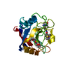 7ogiC 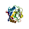 7pmtC 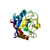 7r2hSC 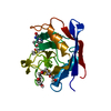 7r2iC 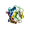 7r2jC 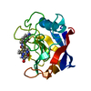 7r2lC C: citing same article ( S: Starting model for refinement |
|---|---|
| Similar structure data |
-Links
 PDBj
PDBj

- Assembly
| Deposited unit | 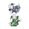
| ||||||||||||
|---|---|---|---|---|---|---|---|---|---|---|---|---|---|
| 1 | 
| ||||||||||||
| 2 | 
| ||||||||||||
| Unit cell |
| ||||||||||||
| Components on special symmetry positions |
|
-Components
| #1: Protein | Mass: 17782.271 Da / Num. of mol.: 2 / Fragment: UNP residues 44-207 / Mutation: K125Q, K133I Source method: isolated from a genetically manipulated source Source: (gene. exp.) Homo sapiens (human) / Gene: PPIF, CYP3 / Plasmid: pet28a / Production host:  #2: Chemical | 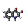  Mass: 191.226 Da / Num. of mol.: 2 / Source method: obtained synthetically / Formula: C11H13NO2 / Feature type: SUBJECT OF INVESTIGATION Mass: 191.226 Da / Num. of mol.: 2 / Source method: obtained synthetically / Formula: C11H13NO2 / Feature type: SUBJECT OF INVESTIGATION#3: Water | ChemComp-HOH / |  Mass: 18.015 Da / Num. of mol.: 444 / Source method: isolated from a natural source / Formula: H2O Mass: 18.015 Da / Num. of mol.: 444 / Source method: isolated from a natural source / Formula: H2OHas ligand of interest | Y | Has protein modification | N | |
|---|
-Experimental details
-Experiment
| Experiment | Method: X-RAY DIFFRACTION / Number of used crystals: 1 |
|---|
- Sample preparation
| Crystal | Density Matthews: 2.66 Å3/Da / Density % sol: 53.7 % |
|---|---|
| Crystal grow | Temperature: 293.15 K / Method: vapor diffusion, sitting drop / pH: 8 / Details: 18 - 23 % PEG3350, 0.1 M TRIS-HCL |
-Data collection
| Diffraction | Mean temperature: 100 K / Serial crystal experiment: N |
|---|---|
| Diffraction source | Source: SYNCHROTRON / Site: ESRF  / Beamline: BM30A / Wavelength: 0.968 Å / Beamline: BM30A / Wavelength: 0.968 Å |
| Detector | Type: DECTRIS EIGER X 4M / Detector: PIXEL / Date: Sep 2, 2018 |
| Radiation | Protocol: SINGLE WAVELENGTH / Monochromatic (M) / Laue (L): M / Scattering type: x-ray |
| Radiation wavelength | Wavelength: 0.968 Å / Relative weight: 1 |
| Reflection | Resolution: 1.549→38.31 Å / Num. obs: 54498 / % possible obs: 98.29 % / Redundancy: 4.4 % / Biso Wilson estimate: 24.04 Å2 / CC1/2: 0.989 / Net I/σ(I): 8.14 |
| Reflection shell | Resolution: 1.549→1.605 Å / Num. unique obs: 5020 / CC1/2: 0.604 |
- Processing
| Software |
| ||||||||||||||||||||||||||||||||||||||||||||||||||||||||||||||||||||||||||||||||||||||||||||||||||||||||||||||||||||||||||||||||||||||||||||
|---|---|---|---|---|---|---|---|---|---|---|---|---|---|---|---|---|---|---|---|---|---|---|---|---|---|---|---|---|---|---|---|---|---|---|---|---|---|---|---|---|---|---|---|---|---|---|---|---|---|---|---|---|---|---|---|---|---|---|---|---|---|---|---|---|---|---|---|---|---|---|---|---|---|---|---|---|---|---|---|---|---|---|---|---|---|---|---|---|---|---|---|---|---|---|---|---|---|---|---|---|---|---|---|---|---|---|---|---|---|---|---|---|---|---|---|---|---|---|---|---|---|---|---|---|---|---|---|---|---|---|---|---|---|---|---|---|---|---|---|---|---|
| Refinement | Method to determine structure: MOLECULAR REPLACEMENT Starting model: 7R2H Resolution: 1.55→38.31 Å / SU ML: 0.2392 / Cross valid method: FREE R-VALUE / σ(F): 1.35 / Phase error: 25.0005 Stereochemistry target values: GeoStd + Monomer Library + CDL v1.2
| ||||||||||||||||||||||||||||||||||||||||||||||||||||||||||||||||||||||||||||||||||||||||||||||||||||||||||||||||||||||||||||||||||||||||||||
| Solvent computation | Shrinkage radii: 0.9 Å / VDW probe radii: 1.11 Å / Solvent model: FLAT BULK SOLVENT MODEL | ||||||||||||||||||||||||||||||||||||||||||||||||||||||||||||||||||||||||||||||||||||||||||||||||||||||||||||||||||||||||||||||||||||||||||||
| Displacement parameters | Biso mean: 28.06 Å2 | ||||||||||||||||||||||||||||||||||||||||||||||||||||||||||||||||||||||||||||||||||||||||||||||||||||||||||||||||||||||||||||||||||||||||||||
| Refinement step | Cycle: LAST / Resolution: 1.55→38.31 Å
| ||||||||||||||||||||||||||||||||||||||||||||||||||||||||||||||||||||||||||||||||||||||||||||||||||||||||||||||||||||||||||||||||||||||||||||
| Refine LS restraints |
| ||||||||||||||||||||||||||||||||||||||||||||||||||||||||||||||||||||||||||||||||||||||||||||||||||||||||||||||||||||||||||||||||||||||||||||
| LS refinement shell |
|
