 Movie
Movie Controller


+ Open data

- Basic information
| Entry | Database: PDB / ID: 7zcl | ||||||
|---|---|---|---|---|---|---|---|
| Title | Unspecific peroxygenase from Collariella virescens | ||||||
 Components Components | (Collariella virescens UPO) x 2 | ||||||
 Keywords Keywords | OXIDOREDUCTASE / Unspecific peroxygenases (UPO) / Collariella virescens / Short UPO family / Molecular structure / Fatty acid epoxidation | ||||||
| Function / homology | Chloroperoxidase / Chloroperoxidase-like / Orthogonal Bundle / Mainly Alpha / HEME C Function and homology information Function and homology information | ||||||
| Biological species |  Collariella virescens (fungus) Collariella virescens (fungus) | ||||||
| Method |  X-RAY DIFFRACTION / SYNCHROTRON / MOLECULAR REPLACEMENT / Resolution: 1.95 Å X-RAY DIFFRACTION / SYNCHROTRON / MOLECULAR REPLACEMENT / Resolution: 1.95 Å | ||||||
 Authors Authors | Santillana, E. / Romero, A. | ||||||
| Funding support |  Spain, 1items Spain, 1items
| ||||||
 Citation Citation | Journal: Antioxidants / Year: 2022 Title: Structural Characterization of Two Short Unspecific Peroxygenases: Two Different Dimeric Arrangements. Authors: Linde, D. / Santillana, E. / Fernandez-Fueyo, E. / Gonzalez-Benjumea, A. / Carro, J. / Gutierrez, A. / Martinez, A.T. / Romero, A. | ||||||
| History |
|
- Structure visualization
| Structure viewer | Molecule: MolmilJmol/JSmol |
|---|
- Downloads & links
-Download
| PDBx/mmCIF format | 7zcl.cif.gz | 137.1 KB | Display | PDBx/mmCIF format |
|---|---|---|---|---|
| PDB format | pdb7zcl.ent.gz | 85.3 KB | Display | PDB format |
| PDBx/mmJSON format | 7zcl.json.gz | Tree view | PDBx/mmJSON format | |
| Others |  Other downloads Other downloads |
-Validation report
| Arichive directory | https://data.pdbj.org/pub/pdb/validation_reports/zc/7zclftp://data.pdbj.org/pub/pdb/validation_reports/zc/7zcl | HTTPS FTP |
|---|
-Related structure data
| Related structure data |  7zbpC 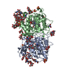 2yorS S: Starting model for refinement C: citing same article ( |
|---|---|
| Similar structure data |
-Links
 PDBj
PDBj


- Assembly
| Deposited unit | 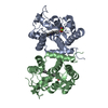
| ||||||||||
|---|---|---|---|---|---|---|---|---|---|---|---|
| 1 |
| ||||||||||
| Unit cell |
|
-Components
| #1: Protein | Mass: 25923.533 Da / Num. of mol.: 1 Source method: isolated from a genetically manipulated source Source: (gene. exp.) Collariella virescens (fungus) / Production host:  | ||||||
|---|---|---|---|---|---|---|---|
| #2: Protein | Mass: 25994.613 Da / Num. of mol.: 1 Source method: isolated from a genetically manipulated source Source: (gene. exp.) Collariella virescens (fungus) / Production host: | ||||||
| #3: Chemical |   Mass: 618.503 Da / Num. of mol.: 2 / Source method: obtained synthetically / Formula: C34H34FeN4O4 / Feature type: SUBJECT OF INVESTIGATION Mass: 618.503 Da / Num. of mol.: 2 / Source method: obtained synthetically / Formula: C34H34FeN4O4 / Feature type: SUBJECT OF INVESTIGATION#4: Chemical |   Mass: 24.305 Da / Num. of mol.: 2 / Source method: obtained synthetically / Formula: Mg Mass: 24.305 Da / Num. of mol.: 2 / Source method: obtained synthetically / Formula: Mg#5: Water | ChemComp-HOH / |  Mass: 18.015 Da / Num. of mol.: 268 / Source method: isolated from a natural source / Formula: H2O Mass: 18.015 Da / Num. of mol.: 268 / Source method: isolated from a natural source / Formula: H2OHas ligand of interest | Y | |
-Experimental details
-Experiment
| Experiment | Method: X-RAY DIFFRACTION / Number of used crystals: 1 |
|---|
- Sample preparation
| Crystal | Density Matthews: 2.24 Å3/Da / Density % sol: 45.2 % |
|---|---|
| Crystal grow | Temperature: 293 K / Method: vapor diffusion, sitting drop / pH: 8.5 Details: 30% PEG 400, 0.1 M Tris, pH 8.5, 0.2 M sodium citrate |
-Data collection
| Diffraction | Mean temperature: 110 K / Serial crystal experiment: N |
|---|---|
| Diffraction source | Source: SYNCHROTRON / Site: ALBA / Beamline: XALOC / Wavelength: 0.97924 Å |
| Detector | Type: DECTRIS PILATUS 6M / Detector: PIXEL / Date: Oct 4, 2020 |
| Radiation | Protocol: SINGLE WAVELENGTH / Monochromatic (M) / Laue (L): M / Scattering type: x-ray |
| Radiation wavelength | Wavelength: 0.97924 Å / Relative weight: 1 |
| Reflection | Resolution: 1.95→70.1 Å / Num. obs: 35859 / % possible obs: 100 % / Redundancy: 8.7 % / Biso Wilson estimate: 22.28 Å2 / Rpim(I) all: 0.057 / Rrim(I) all: 0.191 / Net I/σ(I): 10.1 |
| Reflection shell | Resolution: 1.95→2.06 Å / Redundancy: 8.4 % / Mean I/σ(I) obs: 2.4 / Num. unique obs: 5682 / Rpim(I) all: 0.4 / Rrim(I) all: 0.99 / % possible all: 100 |
- Processing
| Software |
| ||||||||||||||||||||||||||||||||||||||||||||||||||||||||||||||||||||||||||||||||||||||||||||||||||
|---|---|---|---|---|---|---|---|---|---|---|---|---|---|---|---|---|---|---|---|---|---|---|---|---|---|---|---|---|---|---|---|---|---|---|---|---|---|---|---|---|---|---|---|---|---|---|---|---|---|---|---|---|---|---|---|---|---|---|---|---|---|---|---|---|---|---|---|---|---|---|---|---|---|---|---|---|---|---|---|---|---|---|---|---|---|---|---|---|---|---|---|---|---|---|---|---|---|---|---|
| Refinement | Method to determine structure: MOLECULAR REPLACEMENT Starting model: 2YOR Resolution: 1.95→68.47 Å / SU ML: 0.2163 / Cross valid method: FREE R-VALUE / σ(F): 1.36 / Phase error: 23.3086 Stereochemistry target values: GeoStd + Monomer Library + CDL v1.2
| ||||||||||||||||||||||||||||||||||||||||||||||||||||||||||||||||||||||||||||||||||||||||||||||||||
| Solvent computation | Shrinkage radii: 0.9 Å / VDW probe radii: 1.1 Å / Solvent model: FLAT BULK SOLVENT MODEL | ||||||||||||||||||||||||||||||||||||||||||||||||||||||||||||||||||||||||||||||||||||||||||||||||||
| Displacement parameters | Biso mean: 23.54 Å2 | ||||||||||||||||||||||||||||||||||||||||||||||||||||||||||||||||||||||||||||||||||||||||||||||||||
| Refinement step | Cycle: LAST / Resolution: 1.95→68.47 Å
| ||||||||||||||||||||||||||||||||||||||||||||||||||||||||||||||||||||||||||||||||||||||||||||||||||
| Refine LS restraints |
| ||||||||||||||||||||||||||||||||||||||||||||||||||||||||||||||||||||||||||||||||||||||||||||||||||
| LS refinement shell |
|
