 Movie
Movie Controller


+ Open data

- Basic information
| Entry | Database: PDB / ID: 7zbr | ||||||
|---|---|---|---|---|---|---|---|
| Title | Crystal Structure of full-length Bartonella Effector protein 1 | ||||||
 Components Components | Bartonella effector protein (Bep) substrate of VirB T4SS | ||||||
 Keywords Keywords | TOXIN / AMPylation / FIC / T4SS / VirD/B4 / domain insertion / Bartonella / Bartonella effector protein / Bep / BID / Bep intracellular delivery | ||||||
| Function / homology |  Function and homology information Function and homology informationprotein adenylyltransferase / regulation of cell division / nucleotidyltransferase activity Similarity search - Function | ||||||
| Biological species |  Bartonella clarridgeiae (bacteria) Bartonella clarridgeiae (bacteria) | ||||||
| Method |  X-RAY DIFFRACTION / SYNCHROTRON / MOLECULAR REPLACEMENT / Resolution: 3 Å X-RAY DIFFRACTION / SYNCHROTRON / MOLECULAR REPLACEMENT / Resolution: 3 Å | ||||||
 Authors Authors | Huber, M. | ||||||
| Funding support |  Switzerland, 1items Switzerland, 1items
| ||||||
 Citation Citation | Journal: To be published Title: Full-length structure of a FIC effector protein from Bartonella reveals a novel structural domain Authors: Huber, M. / Wagner, A. / Reiners, J. / Seyfert, C.E.M. / Smits, S.H.J. / Schirmer, T. / Dehio, C. | ||||||
| History |
|
- Structure visualization
| Structure viewer | Molecule: MolmilJmol/JSmol |
|---|
- Downloads & links
-Download
| PDBx/mmCIF format | 7zbr.cif.gz | 190.2 KB | Display | PDBx/mmCIF format |
|---|---|---|---|---|
| PDB format | pdb7zbr.ent.gz | 151.3 KB | Display | PDB format |
| PDBx/mmJSON format | 7zbr.json.gz | Tree view | PDBx/mmJSON format | |
| Others |  Other downloads Other downloads |
-Validation report
| Summary document | 7zbr_validation.pdf.gz | 421.7 KB | Display | wwPDB validaton report |
|---|---|---|---|---|
| Full document | 7zbr_full_validation.pdf.gz | 423.9 KB | Display | |
| Data in XML | 7zbr_validation.xml.gz | 18.6 KB | Display | |
| Data in CIF | 7zbr_validation.cif.gz | 24.7 KB | Display | |
| Arichive directory | https://data.pdbj.org/pub/pdb/validation_reports/zb/7zbrftp://data.pdbj.org/pub/pdb/validation_reports/zb/7zbr | HTTPS FTP |
-Related structure data

-Links
 PDBj
PDBj- Assembly
| Deposited unit | 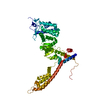
| ||||||||
|---|---|---|---|---|---|---|---|---|---|
| 1 |
| ||||||||
| Unit cell |
|
-Components
| #1: Protein | Mass: 63824.180 Da / Num. of mol.: 1 / Fragment: full-length Source method: isolated from a genetically manipulated source Source: (gene. exp.) Bartonella clarridgeiae (bacteria) / Strain: CIP 104772 / 73 / Gene: BARCL_0069 / Plasmid: pET26 derived / Production host: |
|---|
-Experimental details
-Experiment
| Experiment | Method: X-RAY DIFFRACTION / Number of used crystals: 1 |
|---|
- Sample preparation
| Crystal | Density Matthews: 2.4 Å3/Da / Density % sol: 48.84 % |
|---|---|
| Crystal grow | Temperature: 293.15 K / Method: vapor diffusion, sitting drop / pH: 7.8 Details: 100 mM Hepes pH 7.8, 0.175 mM LiCl and 20% v/v PEG 8000 |
-Data collection
| Diffraction | Mean temperature: 100 K / Serial crystal experiment: N |
|---|---|
| Diffraction source | Source: SYNCHROTRON / Site: SLS / Beamline: X06SA / Wavelength: 1 Å |
| Detector | Type: DECTRIS EIGER X 16M / Detector: PIXEL / Date: Oct 31, 2018 |
| Radiation | Protocol: SINGLE WAVELENGTH / Monochromatic (M) / Laue (L): M / Scattering type: x-ray |
| Radiation wavelength | Wavelength: 1 Å / Relative weight: 1 |
| Reflection | Resolution: 3→46.38 Å / Num. obs: 13003 / % possible obs: 99.63 % / Redundancy: 12.8 % / Biso Wilson estimate: 110.18 Å2 / CC1/2: 0.993 / CC star: 0.998 / Net I/σ(I): 8.1 |
| Reflection shell | Resolution: 3→3.06 Å / Num. unique obs: 1271 / CC1/2: 0.335 |
- Processing
| Software |
| ||||||||||||||||||||||||||||||||||||||||||||||||||||||||||||||||||||||||||||||||||||||||||||||||||||||||||||||||||||||||||||||
|---|---|---|---|---|---|---|---|---|---|---|---|---|---|---|---|---|---|---|---|---|---|---|---|---|---|---|---|---|---|---|---|---|---|---|---|---|---|---|---|---|---|---|---|---|---|---|---|---|---|---|---|---|---|---|---|---|---|---|---|---|---|---|---|---|---|---|---|---|---|---|---|---|---|---|---|---|---|---|---|---|---|---|---|---|---|---|---|---|---|---|---|---|---|---|---|---|---|---|---|---|---|---|---|---|---|---|---|---|---|---|---|---|---|---|---|---|---|---|---|---|---|---|---|---|---|---|---|
| Refinement | Method to determine structure: MOLECULAR REPLACEMENT Starting model: 4NPS, 4YK3 Resolution: 3→46.38 Å / SU ML: 0.6 / Cross valid method: THROUGHOUT / σ(F): 1.33 / Phase error: 39.67 / Stereochemistry target values: ML
| ||||||||||||||||||||||||||||||||||||||||||||||||||||||||||||||||||||||||||||||||||||||||||||||||||||||||||||||||||||||||||||||
| Solvent computation | Shrinkage radii: 0.9 Å / VDW probe radii: 1.11 Å / Solvent model: FLAT BULK SOLVENT MODEL | ||||||||||||||||||||||||||||||||||||||||||||||||||||||||||||||||||||||||||||||||||||||||||||||||||||||||||||||||||||||||||||||
| Displacement parameters | Biso max: 114.43 Å2 / Biso mean: 103.3999 Å2 / Biso min: 96.38 Å2 | ||||||||||||||||||||||||||||||||||||||||||||||||||||||||||||||||||||||||||||||||||||||||||||||||||||||||||||||||||||||||||||||
| Refinement step | Cycle: final / Resolution: 3→46.38 Å
| ||||||||||||||||||||||||||||||||||||||||||||||||||||||||||||||||||||||||||||||||||||||||||||||||||||||||||||||||||||||||||||||
| LS refinement shell | Refine-ID: X-RAY DIFFRACTION / Rfactor Rfree error: 0 / Total num. of bins used: 17
|
