プロトコル: SINGLE WAVELENGTH / 単色(M)・ラウエ(L): M / 散乱光タイプ: x-ray
放射波長
波長: 0.9184 Å / 相対比: 1
反射
解像度: 2→46.42 Å / Num. obs: 91690 / % possible obs: 99.3 % / 冗長度: 6.92 % / Biso Wilson estimate: 33.66 Å2 / CC1/2: 0.997 / Rrim(I) all: 0.167 / Net I/σ(I): 9.1
反射 シェル
解像度: 2→2.12 Å / Num. unique obs: 14692 / CC1/2: 0.773 / % possible all: 98.6
-
位相決定
位相決定
手法: 分子置換
Phasing MR
R rigid body: 0.512
最高解像度
最低解像度
Rotation
46.13 Å
2.64 Å
-
解析
ソフトウェア
名称
バージョン
分類
NB
XDS
データ削減
XSCALE
データスケーリング
MOLREP
位相決定
REFMAC
5.8.0267
精密化
PDB_EXTRACT
3.27
データ抽出
精密化
構造決定の手法: 分子置換 開始モデル: MrBUMP 解像度: 2→46.42 Å / Cor.coef. Fo:Fc: 0.953 / Cor.coef. Fo:Fc free: 0.914 / SU B: 5.983 / SU ML: 0.16 / SU R Cruickshank DPI: 0.2259 / 交差検証法: THROUGHOUT / σ(F): 0 / ESU R: 0.226 / ESU R Free: 0.193 / 立体化学のターゲット値: MAXIMUM LIKELIHOOD 詳細: HYDROGENS HAVE BEEN ADDED IN THE RIDING POSITIONS U VALUES : REFINED INDIVIDUALLY
Rfactor
反射数
%反射
Selection details
Rfree
0.2496
2100
2.3 %
RANDOM
Rwork
0.187
-
-
-
obs
0.1884
89590
99.38 %
-
溶媒の処理
イオンプローブ半径: 0.8 Å / 減衰半径: 0.8 Å / VDWプローブ半径: 1.2 Å / 溶媒モデル: MASK
 ムービー
ムービー コントローラー


 データを開く
 基本情報
基本情報 要素
要素 キーワード
キーワード 機能・相同性情報
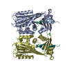
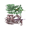
 Ixodes ricinus (ダニ)
 X線回折 /
  データ登録者
 チェコ, 2件
  引用
引用 構造の表示
構造の表示 ダウンロードとリンク
 その他のダウンロード
 PDBj
PDBj
 集合体
集合体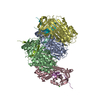


 分子量: 35.453 Da / 分子数: 4 / 由来タイプ: 合成 / 式: Cl
 分子量: 18.015 Da / 分子数: 1276 / 由来タイプ: 天然 / 式: H2O
 試料調製
試料調製 / ビームライン: 14.2 / 波長: 0.9184 Å
 解析
解析