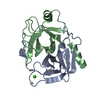
[English] 日本語
Yorodumi
- PDB-7z9f: Human anionic trypsin after autoproteolysis at Arg122 -

+
Open data


ID or keywords:

Loading...

-
Basic information

Entry
Database: PDB / ID: 7z9f
TitleHuman anionic trypsin after autoproteolysis at Arg122
Components(Trypsin-2) x 2
KeywordsHYDROLASE / TRY2 / serine protease / autoproteolysis / digestion
Function / homology
Function and homology information


antimicrobial humoral response / Alpha-defensins / Antimicrobial peptides / Activation of Matrix Metalloproteinases / Collagen degradation / positive regulation of cell adhesion / collagen catabolic process / trypsin / extracellular matrix disassembly / digestion ...antimicrobial humoral response / Alpha-defensins / Antimicrobial peptides / Activation of Matrix Metalloproteinases / Collagen degradation / positive regulation of cell adhesion / collagen catabolic process / trypsin / extracellular matrix disassembly / digestion / serine-type peptidase activity / extracellular matrix / metalloendopeptidase activity / azurophil granule lumen / positive regulation of cell growth / serine-type endopeptidase activity / calcium ion binding / Neutrophil degranulation / proteolysis / extracellular space / extracellular region
Similarity search - Function
Serine proteases, trypsin family, histidine active site / Serine proteases, trypsin family, serine active site / Peptidase S1A, chymotrypsin family / Serine proteases, trypsin family, histidine active site. / Serine proteases, trypsin domain profile. / Serine proteases, trypsin family, serine active site. / Trypsin-like serine protease / Serine proteases, trypsin domain / Trypsin / Peptidase S1, PA clan, chymotrypsin-like fold / Peptidase S1, PA clan
Similarity search - Domain/homology
Biological speciesHomo sapiens (human)
MethodX-RAY DIFFRACTION / SYNCHROTRON / MOLECULAR REPLACEMENT / Resolution: 1.7 Å
AuthorsNagel, F. / Palm, G.J. / Delcea, M. / Lammers, M.
Funding supportEuropean Union, 1items
OrganizationGrant numberCountry
European Research Council (ERC)637877European Union
CitationJournal: J Inflamm Res / Year: 2022
Title: Structural Basis of the Pancreatitis-Associated Autoproteolytic Failsafe Mechanism in Human Anionic Trypsin.
Authors: Nagel, F. / Susemihl, A. / Geist, N. / Mohlis, K. / Palm, G.J. / Lammers, M. / Delcea, M.
History
DepositionMar 21, 2022Deposition site: PDBE / Processing site: PDBE
Revision 1.0Jun 29, 2022Provider: repository / Type: Initial release
Revision 1.1Jul 13, 2022Group: Database references / Category: citation / citation_author
Item: _citation.journal_id_ISSN / _citation.journal_volume ..._citation.journal_id_ISSN / _citation.journal_volume / _citation.page_first / _citation.page_last / _citation.pdbx_database_id_PubMed / _citation.title / _citation_author.identifier_ORCID / _citation_author.name
Revision 1.2May 1, 2024Group: Data collection / Database references / Refinement description
Category: chem_comp_atom / chem_comp_bond ...chem_comp_atom / chem_comp_bond / citation / pdbx_initial_refinement_model
Item: _citation.country

-
Structure visualization

Structure viewerMolecule:
MolmilJmol/JSmol

Downloads & links

-
Assembly

Deposited unit
X: Trypsin-2
A: Trypsin-2
Y: Trypsin-2
B: Trypsin-2
hetero molecules


Theoretical massNumber of molelcules
Total (without water)48,2386
Polymers48,1584
Non-polymers802
Water13,872770
1
X: Trypsin-2
Y: Trypsin-2
hetero molecules


Theoretical massNumber of molelcules
Total (without water)24,1193
Polymers24,0792
Non-polymers401
Water362
TypeNameSymmetry operationNumber
identity operation1_555x,y,z1
Buried area4390 Å2
ΔGint-43 kcal/mol
Surface area9820 Å2
2


  • Idetical with deposited unit
  • defined by author
  • Evidence: gel filtration
TypeNameSymmetry operationNumber
identity operation1_555x,y,z1
Buried area10450 Å2
ΔGint-85 kcal/mol
Surface area17870 Å2
3
A: Trypsin-2
B: Trypsin-2
hetero molecules


Theoretical massNumber of molelcules
Total (without water)24,1193
Polymers24,0792
Non-polymers401
Water362
TypeNameSymmetry operationNumber
identity operation1_555x,y,z1
Buried area4380 Å2
ΔGint-43 kcal/mol
Surface area9730 Å2
Unit cell
Length a, b, c (Å)59.060, 80.790, 87.940
Angle α, β, γ (deg.)90.000, 90.000, 90.000
Int Tables number19
Space group name H-MP212121
Space group name HallP2ac2ab
Symmetry operation#1: x,y,z
#2: x+1/2,-y+1/2,-z
#3: -x,y+1/2,-z+1/2
#4: -x+1/2,-y,z+1/2

-
Components

#1: Protein Trypsin-2 / Anionic trypsinogen / Serine protease 2 / Trypsin II


Mass: 11072.527 Da / Num. of mol.: 2
Source method: isolated from a genetically manipulated source
Source: (gene. exp.) Homo sapiens (human) / Gene: PRSS2, TRY2, TRYP2 / Plasmid: pET47b / Production host: Escherichia coli BL21(DE3) (bacteria) / References: UniProt: P07478, trypsin
#2: Protein Trypsin-2 / Anionic trypsinogen / Serine protease 2 / Trypsin II


Mass: 13006.325 Da / Num. of mol.: 2
Source method: isolated from a genetically manipulated source
Source: (gene. exp.) Homo sapiens (human) / Gene: PRSS2, TRY2, TRYP2 / Plasmid: pET47b / Production host: Escherichia coli BL21(DE3) (bacteria) / References: UniProt: P07478, trypsin
#3: Chemical ChemComp-CA / CALCIUM ION


Mass: 40.078 Da / Num. of mol.: 2 / Source method: obtained synthetically / Formula: Ca / Feature type: SUBJECT OF INVESTIGATION
#4: Water ChemComp-HOH / water


Mass: 18.015 Da / Num. of mol.: 770 / Source method: isolated from a natural source / Formula: H2O
Has ligand of interestY

-
Experimental details

-
Experiment

ExperimentMethod: X-RAY DIFFRACTION / Number of used crystals: 1

-
Sample preparation

CrystalDensity Matthews: 2.18 Å3/Da / Density % sol: 43.62 %
Crystal growTemperature: 293 K / Method: vapor diffusion, hanging drop / pH: 7.5
Details: 1 uL protein (15 mg/mL in 20 mM Hepes pH 7.4, 150 mM NaCl, 1 mM CaCl2) + 1 uL well solution (22% PEG 4000, 0.1 M Hepes pH 7.5, 0.1 M NaAc)

-
Data collection

DiffractionMean temperature: 100 K / Serial crystal experiment: N
Diffraction sourceSource: SYNCHROTRON / Site: BESSY / Beamline: 14.2 / Wavelength: 0.9184 Å
DetectorType: DECTRIS PILATUS3 S 2M / Detector: PIXEL / Date: Dec 9, 2021 / Details: DCM Si(111)
RadiationMonochromator: DCM Si(111) / Protocol: SINGLE WAVELENGTH / Monochromatic (M) / Laue (L): M / Scattering type: x-ray
Radiation wavelengthWavelength: 0.9184 Å / Relative weight: 1
ReflectionResolution: 1.7→50 Å / Num. obs: 47119 / % possible obs: 99.6 % / Observed criterion σ(I): -3 / Redundancy: 12.9 % / Biso Wilson estimate: 16.59 Å2 / CC1/2: 0.999 / Rrim(I) all: 0.169 / Rsym value: 0.162 / Net I/σ(I): 11.9
Reflection shellResolution: 1.7→1.8 Å / Redundancy: 12.8 % / Mean I/σ(I) obs: 2.03 / Num. unique obs: 7342 / CC1/2: 0.82 / Rrim(I) all: 1.252 / Rsym value: 1.203 / % possible all: 97.3

-
Processing

Software
NameVersionClassification
PHENIX1.18.2_3874refinement
XDSVERSION Feb 5, 2021data reduction
XDSVERSION Feb 5, 2021data scaling
PHASER2.8.3phasing
RefinementMethod to determine structure: MOLECULAR REPLACEMENT
Starting model: 1TRN_A

Resolution: 1.7→47.68 Å / SU ML: 0.1549 / Cross valid method: FREE R-VALUE / σ(F): 1.35 / Phase error: 23.3629
Stereochemistry target values: GeoStd + Monomer Library + CDL v1.2
RfactorNum. reflection% reflection
Rfree0.2186 1516 3.22 %
Rwork0.187 45524 -
obs0.188 47040 99.4 %
Solvent computationShrinkage radii: 0.9 Å / VDW probe radii: 1.11 Å / Solvent model: FLAT BULK SOLVENT MODEL
Displacement parametersBiso mean: 20.03 Å2
Refinement stepCycle: LAST / Resolution: 1.7→47.68 Å
ProteinNucleic acidLigandSolventTotal
Num. atoms3368 0 2 770 4140
Refine LS restraints
Refine-IDTypeDev idealNumber
X-RAY DIFFRACTIONf_bond_d0.00643440
X-RAY DIFFRACTIONf_angle_d0.90234678
X-RAY DIFFRACTIONf_chiral_restr0.0642512
X-RAY DIFFRACTIONf_plane_restr0.0061616
X-RAY DIFFRACTIONf_dihedral_angle_d17.7046478
LS refinement shell
Resolution (Å)Rfactor RfreeNum. reflection RfreeRfactor RworkNum. reflection RworkRefine-ID% reflection obs (%)
1.7-1.750.26331300.23113902X-RAY DIFFRACTION95.09
1.75-1.810.27511350.22644066X-RAY DIFFRACTION99.95
1.81-1.890.27771380.22224132X-RAY DIFFRACTION99.98
1.89-1.970.25381370.21094113X-RAY DIFFRACTION99.77
1.97-2.080.22421370.19584119X-RAY DIFFRACTION99.91
2.08-2.210.21161370.18464130X-RAY DIFFRACTION99.84
2.21-2.380.21531390.18814149X-RAY DIFFRACTION99.79
2.38-2.620.21171370.18924126X-RAY DIFFRACTION99.84
2.62-2.990.23441380.18954186X-RAY DIFFRACTION99.93
2.99-3.770.1871410.16784220X-RAY DIFFRACTION99.84
3.77-47.680.20621470.17194381X-RAY DIFFRACTION99.43
Refinement TLS params.Method: refined / Origin x: -1.81458986763 Å / Origin y: 0.0222217079109 Å / Origin z: 8.6268521355 Å
111213212223313233
T0.100686823161 Å20.0168593554767 Å20.00524151772626 Å2-0.0834242969554 Å20.00534738229264 Å2--0.0876909091827 Å2
L0.38230959358 °20.0803998581143 °20.0695177137913 °2-0.189084145344 °20.0979027523142 °2--0.195212649796 °2
S-0.0192224983671 Å °-0.00479364644667 Å °-0.00510296173351 Å °0.00402497463132 Å °0.0101878294224 Å °0.0126340930777 Å °-0.0278423810289 Å °0.00465508084958 Å °0.000159665955469 Å °
Refinement TLS groupSelection details: all

+
About Yorodumi

-
News

-
Feb 9, 2022. New format data for meta-information of EMDB entries

New format data for meta-information of EMDB entries

  • Version 3 of the EMDB header file is now the official format.
  • The previous official version 1.9 will be removed from the archive.

Related info.:EMDB header

External links:wwPDB to switch to version 3 of the EMDB data model

-
Aug 12, 2020. Covid-19 info

Covid-19 info

URL: https://pdbjlvh1.pdbj.org/emnavi/covid19.php

New page: Covid-19 featured information page in EM Navigator.

Related info.:Covid-19 info / Mar 5, 2020. Novel coronavirus structure data

+
Mar 5, 2020. Novel coronavirus structure data

Novel coronavirus structure data

Related info.:Yorodumi Speices / Aug 12, 2020. Covid-19 info

External links:COVID-19 featured content - PDBj / Molecule of the Month (242):Coronavirus Proteases

+
Jan 31, 2019. EMDB accession codes are about to change! (news from PDBe EMDB page)

EMDB accession codes are about to change! (news from PDBe EMDB page)

  • The allocation of 4 digits for EMDB accession codes will soon come to an end. Whilst these codes will remain in use, new EMDB accession codes will include an additional digit and will expand incrementally as the available range of codes is exhausted. The current 4-digit format prefixed with “EMD-” (i.e. EMD-XXXX) will advance to a 5-digit format (i.e. EMD-XXXXX), and so on. It is currently estimated that the 4-digit codes will be depleted around Spring 2019, at which point the 5-digit format will come into force.
  • The EM Navigator/Yorodumi systems omit the EMD- prefix.

Related info.:Q: What is EMD? / ID/Accession-code notation in Yorodumi/EM Navigator

External links:EMDB Accession Codes are Changing Soon! / Contact to PDBj

+
Jul 12, 2017. Major update of PDB

Major update of PDB

  • wwPDB released updated PDB data conforming to the new PDBx/mmCIF dictionary.
  • This is a major update changing the version number from 4 to 5, and with Remediation, in which all the entries are updated.
  • In this update, many items about electron microscopy experimental information are reorganized (e.g. em_software).
  • Now, EM Navigator and Yorodumi are based on the updated data.

External links:wwPDB Remediation / Enriched Model Files Conforming to OneDep Data Standards Now Available in the PDB FTP Archive

-
Yorodumi

Thousand views of thousand structures

  • Yorodumi is a browser for structure data from EMDB, PDB, SASBDB, etc.
  • This page is also the successor to EM Navigator detail page, and also detail information page/front-end page for Omokage search.
  • The word "yorodu" (or yorozu) is an old Japanese word meaning "ten thousand". "mi" (miru) is to see.

Related info.:EMDB / PDB / SASBDB / Comparison of 3 databanks / Yorodumi Search / Aug 31, 2016. New EM Navigator & Yorodumi / Yorodumi Papers / Jmol/JSmol / Function and homology information / Changes in new EM Navigator and Yorodumi

Read more