 Movie
Movie Controller

[English] 日本語
 Yorodumi
- PDB-7z98: Crystal structure of F191M variant Variovorax paradoxus indole mo... -
+ Open data

- Basic information
| Entry | Database: PDB / ID: 7z98 | ||||||
|---|---|---|---|---|---|---|---|
| Title | Crystal structure of F191M variant Variovorax paradoxus indole monooxygenase (VpIndA1) in complex with methyl phenyl sulfide | ||||||
 Components Components | Putative dehydrogenase/oxygenase subunit (Flavoprotein) | ||||||
 Keywords Keywords | OXIDOREDUCTASE / FAD-dependent monooxygenase / styrene monooxygenase / StyA1 / IMO | ||||||
| Function / homology | Styrene monooxygenase StyA, putative substrate binding domain / Styrene monooxygenase A putative substrate binding domain / FAD/NAD(P)-binding domain superfamily / nucleotide binding / (methylsulfanyl)benzene / FLAVIN-ADENINE DINUCLEOTIDE / Putative dehydrogenase/oxygenase subunit (Flavoprotein) Function and homology information Function and homology information | ||||||
| Biological species |  Variovorax paradoxus EPS (bacteria) Variovorax paradoxus EPS (bacteria) | ||||||
| Method |  X-RAY DIFFRACTION / SYNCHROTRON / FOURIER SYNTHESIS / Resolution: 1.73 Å X-RAY DIFFRACTION / SYNCHROTRON / FOURIER SYNTHESIS / Resolution: 1.73 Å | ||||||
 Authors Authors | Kratky, J. / Weisse, R. / Strater, N. | ||||||
| Funding support | 1items
| ||||||
 Citation Citation | Journal: Angew.Chem.Int.Ed.Engl. / Year: 2023 Title: Structural and Mechanistic Studies on Substrate and Stereoselectivity of the Indole Monooxygenase VpIndA1: New Avenues for Biocatalytic Epoxidations and Sulfoxidations. Authors: Kratky, J. / Eggerichs, D. / Heine, T. / Hofmann, S. / Sowa, P. / Weisse, R.H. / Tischler, D. / Strater, N. | ||||||
| History |
|
- Structure visualization
| Structure viewer | Molecule: MolmilJmol/JSmol |
|---|
- Downloads & links
-Download
| PDBx/mmCIF format | 7z98.cif.gz | 174.6 KB | Display | PDBx/mmCIF format |
|---|---|---|---|---|
| PDB format | pdb7z98.ent.gz | 136.6 KB | Display | PDB format |
| PDBx/mmJSON format | 7z98.json.gz | Tree view | PDBx/mmJSON format | |
| Others |  Other downloads Other downloads |
-Validation report
| Arichive directory | https://data.pdbj.org/pub/pdb/validation_reports/z9/7z98ftp://data.pdbj.org/pub/pdb/validation_reports/z9/7z98 | HTTPS FTP |
|---|
-Related structure data
| Related structure data |  7z4xSC  7z94C 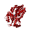 7z97C 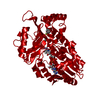 7z99C 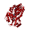 7zcaC S: Starting model for refinement C: citing same article ( |
|---|---|
| Similar structure data |
-Links
 PDBj
PDBj- Assembly
| Deposited unit | 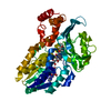
| ||||||||
|---|---|---|---|---|---|---|---|---|---|
| 1 | 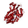
| ||||||||
| Unit cell |
|
-Components
| #1: Protein | Mass: 47308.590 Da / Num. of mol.: 1 Source method: isolated from a genetically manipulated source Source: (gene. exp.) Variovorax paradoxus EPS (bacteria) / Strain: EPS / Gene: Varpa_4903 / Production host: | ||||||
|---|---|---|---|---|---|---|---|
| #2: Chemical | ChemComp-FAD /   Mass: 785.550 Da / Num. of mol.: 1 / Source method: obtained synthetically / Formula: C27H33N9O15P2 / Comment: FAD*YM Mass: 785.550 Da / Num. of mol.: 1 / Source method: obtained synthetically / Formula: C27H33N9O15P2 / Comment: FAD*YM | ||||||
| #3: Chemical |   Mass: 62.068 Da / Num. of mol.: 2 / Source method: obtained synthetically / Formula: C2H6O2 Mass: 62.068 Da / Num. of mol.: 2 / Source method: obtained synthetically / Formula: C2H6O2#4: Chemical | ChemComp-16R / ( | 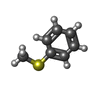  Mass: 124.203 Da / Num. of mol.: 1 / Source method: obtained synthetically / Formula: C7H8S / Feature type: SUBJECT OF INVESTIGATION Mass: 124.203 Da / Num. of mol.: 1 / Source method: obtained synthetically / Formula: C7H8S / Feature type: SUBJECT OF INVESTIGATION#5: Water | ChemComp-HOH / |  Mass: 18.015 Da / Num. of mol.: 367 / Source method: isolated from a natural source / Formula: H2O Mass: 18.015 Da / Num. of mol.: 367 / Source method: isolated from a natural source / Formula: H2OHas ligand of interest | Y | |
-Experimental details
-Experiment
| Experiment | Method: X-RAY DIFFRACTION / Number of used crystals: 1 |
|---|
- Sample preparation
| Crystal | Density Matthews: 3.02 Å3/Da / Density % sol: 59.32 % |
|---|---|
| Crystal grow | Temperature: 293 K / Method: vapor diffusion, hanging drop / pH: 5 Details: 100 mM Na-acetate pH 5, 1400 mM Na-malonate, 1 mM DTT, 3 mM FAD, 1 mM methyl phenyl sulfide, cryo: 15 % (v/v) ethylene glycol |
-Data collection
| Diffraction | Mean temperature: 100 K / Serial crystal experiment: N | ||||||||||||||||||||||||||||||
|---|---|---|---|---|---|---|---|---|---|---|---|---|---|---|---|---|---|---|---|---|---|---|---|---|---|---|---|---|---|---|---|
| Diffraction source | Source: SYNCHROTRON / Site: BESSY  / Beamline: 14.1 / Wavelength: 0.9184 Å / Beamline: 14.1 / Wavelength: 0.9184 Å | ||||||||||||||||||||||||||||||
| Detector | Type: DECTRIS PILATUS 6M / Detector: PIXEL / Date: Jan 23, 2018 | ||||||||||||||||||||||||||||||
| Radiation | Protocol: SINGLE WAVELENGTH / Monochromatic (M) / Laue (L): M / Scattering type: x-ray | ||||||||||||||||||||||||||||||
| Radiation wavelength | Wavelength: 0.9184 Å / Relative weight: 1 | ||||||||||||||||||||||||||||||
| Reflection | Resolution: 1.73→47.24 Å / Num. obs: 59530 / % possible obs: 98.2 % / Redundancy: 8.6 % / CC1/2: 0.999 / Rmerge(I) obs: 0.085 / Rpim(I) all: 0.03 / Rrim(I) all: 0.09 / Net I/σ(I): 12.4 / Num. measured all: 514810 | ||||||||||||||||||||||||||||||
| Reflection shell | Diffraction-ID: 1
|
- Processing
| Software |
| |||||||||||||||||||||||||||||||||||||||||||||||||||||||||||||||||||||||||||||||||||||||||||||||||||||||||||||||||||||||||||||||||||||||||||||||||||||||||||||||||
|---|---|---|---|---|---|---|---|---|---|---|---|---|---|---|---|---|---|---|---|---|---|---|---|---|---|---|---|---|---|---|---|---|---|---|---|---|---|---|---|---|---|---|---|---|---|---|---|---|---|---|---|---|---|---|---|---|---|---|---|---|---|---|---|---|---|---|---|---|---|---|---|---|---|---|---|---|---|---|---|---|---|---|---|---|---|---|---|---|---|---|---|---|---|---|---|---|---|---|---|---|---|---|---|---|---|---|---|---|---|---|---|---|---|---|---|---|---|---|---|---|---|---|---|---|---|---|---|---|---|---|---|---|---|---|---|---|---|---|---|---|---|---|---|---|---|---|---|---|---|---|---|---|---|---|---|---|---|---|---|---|---|---|
| Refinement | Method to determine structure: FOURIER SYNTHESIS Starting model: 7z4x Resolution: 1.73→45.93 Å / SU ML: 0.27 / Cross valid method: THROUGHOUT / σ(F): 1.34 / Phase error: 26.11 / Stereochemistry target values: ML
| |||||||||||||||||||||||||||||||||||||||||||||||||||||||||||||||||||||||||||||||||||||||||||||||||||||||||||||||||||||||||||||||||||||||||||||||||||||||||||||||||
| Solvent computation | Shrinkage radii: 0.9 Å / VDW probe radii: 1.11 Å / Solvent model: FLAT BULK SOLVENT MODEL | |||||||||||||||||||||||||||||||||||||||||||||||||||||||||||||||||||||||||||||||||||||||||||||||||||||||||||||||||||||||||||||||||||||||||||||||||||||||||||||||||
| Displacement parameters | Biso max: 118.52 Å2 / Biso mean: 37.6668 Å2 / Biso min: 18.01 Å2 | |||||||||||||||||||||||||||||||||||||||||||||||||||||||||||||||||||||||||||||||||||||||||||||||||||||||||||||||||||||||||||||||||||||||||||||||||||||||||||||||||
| Refinement step | Cycle: final / Resolution: 1.73→45.93 Å
| |||||||||||||||||||||||||||||||||||||||||||||||||||||||||||||||||||||||||||||||||||||||||||||||||||||||||||||||||||||||||||||||||||||||||||||||||||||||||||||||||
| LS refinement shell | Refine-ID: X-RAY DIFFRACTION / Rfactor Rfree error: 0 / Total num. of bins used: 22
|
