 Movie
Movie Controller

[English] 日本語
 Yorodumi
- PDB-7z6n: Crystal structure of Zn2+-transporter BbZIP in a metal-stripped state -
+ Open data

- Basic information
| Entry | Database: PDB / ID: 7z6n | ||||||
|---|---|---|---|---|---|---|---|
| Title | Crystal structure of Zn2+-transporter BbZIP in a metal-stripped state | ||||||
 Components Components | Putative membrane protein | ||||||
 Keywords Keywords | MEMBRANE PROTEIN / Zinc transporters / Zrt/Irt-like proteins (ZIPs) / solute carrier 39 (SLC39) family / BbZIP / transport mechanism / elevator mechanism | ||||||
| Function / homology | Zinc/iron permease / ZIP Zinc transporter / zinc ion transmembrane transporter activity / plasma membrane / Zinc transporter ZIPB Function and homology information Function and homology information | ||||||
| Biological species |  Bordetella bronchiseptica (bacteria) Bordetella bronchiseptica (bacteria) | ||||||
| Method |  X-RAY DIFFRACTION / SYNCHROTRON / MOLECULAR REPLACEMENT / Resolution: 2.57 Å X-RAY DIFFRACTION / SYNCHROTRON / MOLECULAR REPLACEMENT / Resolution: 2.57 Å | ||||||
 Authors Authors | Wiuf, A. / Steffen, J.H. / Becares, E.R. / Groenberg, C. / Mahato, D.R. / Rasmussen, S.G.F. / Andersson, M. / Croll, T. / Gotfryd, K. / Gourdon, P. | ||||||
| Funding support |  Denmark, 1items Denmark, 1items
| ||||||
 Citation Citation | Journal: Sci Adv / Year: 2022 Title: The two-domain elevator-type mechanism of zinc-transporting ZIP proteins. Authors: Wiuf, A. / Steffen, J.H. / Becares, E.R. / Gronberg, C. / Mahato, D.R. / Rasmussen, S.G.F. / Andersson, M. / Croll, T. / Gotfryd, K. / Gourdon, P. | ||||||
| History |
|
- Structure visualization
| Structure viewer | Molecule: MolmilJmol/JSmol |
|---|
- Downloads & links
-Download
| PDBx/mmCIF format | 7z6n.cif.gz | 129.8 KB | Display | PDBx/mmCIF format |
|---|---|---|---|---|
| PDB format | pdb7z6n.ent.gz | 81.5 KB | Display | PDB format |
| PDBx/mmJSON format | 7z6n.json.gz | Tree view | PDBx/mmJSON format | |
| Others |  Other downloads Other downloads |
-Validation report
| Arichive directory | https://data.pdbj.org/pub/pdb/validation_reports/z6/7z6nftp://data.pdbj.org/pub/pdb/validation_reports/z6/7z6n | HTTPS FTP |
|---|
-Related structure data
| Related structure data |  7z6mC 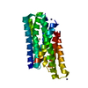 5tsaS S: Starting model for refinement C: citing same article ( |
|---|---|
| Similar structure data |
-Links
 PDBj
PDBj- Assembly
| Deposited unit | 
| ||||||||||||
|---|---|---|---|---|---|---|---|---|---|---|---|---|---|
| 1 |
| ||||||||||||
| Unit cell |
| ||||||||||||
| Components on special symmetry positions |
|
-Components
| #1: Protein | Mass: 33180.648 Da / Num. of mol.: 2 Source method: isolated from a genetically manipulated source Source: (gene. exp.) Bordetella bronchiseptica (bacteria) / Strain: ATCC BAA-588 / NCTC 13252 / RB50 / Gene: BB2405 / Plasmid: pET15b+ / Production host: #2: Chemical | ChemComp-SO4 /   Mass: 96.063 Da / Num. of mol.: 6 / Source method: obtained synthetically / Formula: SO4 Mass: 96.063 Da / Num. of mol.: 6 / Source method: obtained synthetically / Formula: SO4#3: Water | ChemComp-HOH / |  Mass: 18.015 Da / Num. of mol.: 10 / Source method: isolated from a natural source / Formula: H2O Mass: 18.015 Da / Num. of mol.: 10 / Source method: isolated from a natural source / Formula: H2OHas ligand of interest | N | |
|---|
-Experimental details
-Experiment
| Experiment | Method: X-RAY DIFFRACTION / Number of used crystals: 1 |
|---|
- Sample preparation
| Crystal | Density Matthews: 2.79 Å3/Da / Density % sol: 55.94 % |
|---|---|
| Crystal grow | Temperature: 291 K / Method: lipidic cubic phase / pH: 4.5 Details: 100 mM NaOAc-HOAc pH = 4.5, 100-400 mM LiSO4 and 25-40 % PEG400 |
-Data collection
| Diffraction | Mean temperature: 100 K / Serial crystal experiment: N |
|---|---|
| Diffraction source | Source: SYNCHROTRON / Site: SLS  / Beamline: X06SA / Wavelength: 1 Å / Beamline: X06SA / Wavelength: 1 Å |
| Detector | Type: DECTRIS EIGER R 4M / Detector: PIXEL / Date: Aug 22, 2019 |
| Radiation | Protocol: SINGLE WAVELENGTH / Monochromatic (M) / Laue (L): M / Scattering type: x-ray |
| Radiation wavelength | Wavelength: 1 Å / Relative weight: 1 |
| Reflection | Resolution: 2.57→41.17 Å / Num. obs: 22269 / % possible obs: 89.77 % / Redundancy: 15.5 % / CC1/2: 0.999 / CC star: 1 / Rpim(I) all: 0.1222 / Net I/av σ(I): 5.02 / Net I/σ(I): 5.02 |
| Reflection shell | Resolution: 2.57→2.66 Å / Num. unique obs: 663 / CC1/2: 0.0526 / CC star: 0.316 / % possible all: 29.81 |
- Processing
| Software |
| ||||||||||||||||||||||||||||||||||||||||||
|---|---|---|---|---|---|---|---|---|---|---|---|---|---|---|---|---|---|---|---|---|---|---|---|---|---|---|---|---|---|---|---|---|---|---|---|---|---|---|---|---|---|---|---|
| Refinement | Method to determine structure: MOLECULAR REPLACEMENT Starting model: 5TSA Resolution: 2.57→41.17 Å / SU ML: 0.3624 / Cross valid method: FREE R-VALUE / σ(F): 1.34 / Phase error: 41.6506 Stereochemistry target values: GeoStd + Monomer Library + CDL v1.2
| ||||||||||||||||||||||||||||||||||||||||||
| Solvent computation | Shrinkage radii: 0.9 Å / VDW probe radii: 1.11 Å / Solvent model: FLAT BULK SOLVENT MODEL | ||||||||||||||||||||||||||||||||||||||||||
| Refinement step | Cycle: LAST / Resolution: 2.57→41.17 Å
| ||||||||||||||||||||||||||||||||||||||||||
| Refine LS restraints |
| ||||||||||||||||||||||||||||||||||||||||||
| LS refinement shell |
|
