 Movie
Movie Controller

[English] 日本語
 Yorodumi
- PDB-7z5b: Structure of the mouse 8-oxoguanine DNA Glycosylase mOGG1 in comp... -
+ Open data

- Basic information
| Entry | Database: PDB / ID: 7z5b | ||||||
|---|---|---|---|---|---|---|---|
| Title | Structure of the mouse 8-oxoguanine DNA Glycosylase mOGG1 in complex with ligand TH013546 | ||||||
 Components Components | N-glycosylase/DNA lyase | ||||||
 Keywords Keywords | DNA BINDING PROTEIN / OGG1 / glycosylase / inhibitor | ||||||
| Function / homology |  Function and homology information Function and homology informationCleavage of the damaged pyrimidine / oxidized base lesion DNA N-glycosylase activity / Displacement of DNA glycosylase by APEX1 / APEX1-Independent Resolution of AP Sites via the Single Nucleotide Replacement Pathway / Recognition and association of DNA glycosylase with site containing an affected purine / Cleavage of the damaged purine / negative regulation of double-strand break repair via single-strand annealing / oxidized purine nucleobase lesion DNA N-glycosylase activity / DNA N-glycosylase activity / 8-oxo-7,8-dihydroguanine DNA N-glycosylase activity ...Cleavage of the damaged pyrimidine / oxidized base lesion DNA N-glycosylase activity / Displacement of DNA glycosylase by APEX1 / APEX1-Independent Resolution of AP Sites via the Single Nucleotide Replacement Pathway / Recognition and association of DNA glycosylase with site containing an affected purine / Cleavage of the damaged purine / negative regulation of double-strand break repair via single-strand annealing / oxidized purine nucleobase lesion DNA N-glycosylase activity / DNA N-glycosylase activity / 8-oxo-7,8-dihydroguanine DNA N-glycosylase activity / positive regulation of gene expression via chromosomal CpG island demethylation / Hydrolases; Glycosylases; Hydrolysing N-glycosyl compounds / oxidized purine DNA binding / class I DNA-(apurinic or apyrimidinic site) endonuclease activity / DNA-(apurinic or apyrimidinic site) lyase / cellular response to reactive oxygen species / nucleotide-excision repair / response to radiation / base-excision repair / nuclear matrix / response to oxidative stress / microtubule binding / nuclear speck / RNA polymerase II cis-regulatory region sequence-specific DNA binding / DNA repair / DNA damage response / regulation of DNA-templated transcription / enzyme binding / positive regulation of transcription by RNA polymerase II / protein-containing complex / mitochondrion / DNA binding / nucleoplasm / nucleus Similarity search - Function | ||||||
| Biological species |  | ||||||
| Method |  X-RAY DIFFRACTION / SYNCHROTRON / MOLECULAR REPLACEMENT / Resolution: 2.6 Å X-RAY DIFFRACTION / SYNCHROTRON / MOLECULAR REPLACEMENT / Resolution: 2.6 Å | ||||||
 Authors Authors | Scaletti, E. / Stenmark, P. | ||||||
| Funding support |  Sweden, 1items Sweden, 1items
| ||||||
 Citation Citation | Journal: Nat Commun / Year: 2025 Title: Virtual fragment screening for DNA repair inhibitors in vast chemical space. Authors: Luttens, A. / Vo, D.D. / Scaletti, E.R. / Wiita, E. / Almlof, I. / Wallner, O. / Davies, J. / Kosenina, S. / Meng, L. / Long, M. / Mortusewicz, O. / Masuyer, G. / Ballante, F. / Michel, M. / ...Authors: Luttens, A. / Vo, D.D. / Scaletti, E.R. / Wiita, E. / Almlof, I. / Wallner, O. / Davies, J. / Kosenina, S. / Meng, L. / Long, M. / Mortusewicz, O. / Masuyer, G. / Ballante, F. / Michel, M. / Homan, E. / Scobie, M. / Kalderen, C. / Warpman Berglund, U. / Tarnovskiy, A.V. / Radchenko, D.S. / Moroz, Y.S. / Kihlberg, J. / Stenmark, P. / Helleday, T. / Carlsson, J. | ||||||
| History |
|
- Structure visualization
| Structure viewer | Molecule: MolmilJmol/JSmol |
|---|
- Downloads & links
-Download
| PDBx/mmCIF format | 7z5b.cif.gz | 195.4 KB | Display | PDBx/mmCIF format |
|---|---|---|---|---|
| PDB format | pdb7z5b.ent.gz | 154 KB | Display | PDB format |
| PDBx/mmJSON format | 7z5b.json.gz | Tree view | PDBx/mmJSON format | |
| Others |  Other downloads Other downloads |
-Validation report
| Arichive directory | https://data.pdbj.org/pub/pdb/validation_reports/z5/7z5bftp://data.pdbj.org/pub/pdb/validation_reports/z5/7z5b | HTTPS FTP |
|---|
-Related structure data
| Related structure data | 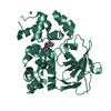 7qelC 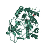 7z3yC 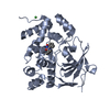 7z5rC 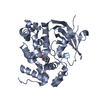 7zc7C 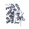 7zg3C 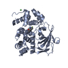 8cexC 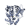 8ceyC 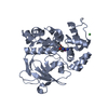 6g3yS S: Starting model for refinement C: citing same article ( |
|---|---|
| Similar structure data |
-Links
 PDBj
PDBj
- Assembly
| Deposited unit | 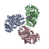
| ||||||||
|---|---|---|---|---|---|---|---|---|---|
| 1 | 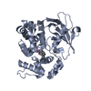
| ||||||||
| 2 | 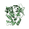
| ||||||||
| 3 | 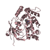
| ||||||||
| Unit cell |
|
-Components
| #1: Protein | Mass: 35816.652 Da / Num. of mol.: 3 Source method: isolated from a genetically manipulated source Source: (gene. exp.)  References: UniProt: O08760, Hydrolases; Glycosylases; Hydrolysing N-glycosyl compounds, DNA-(apurinic or apyrimidinic site) lyase #2: Chemical | 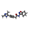  Mass: 350.414 Da / Num. of mol.: 3 / Source method: obtained synthetically / Formula: C20H22N4O2 / Feature type: SUBJECT OF INVESTIGATION Mass: 350.414 Da / Num. of mol.: 3 / Source method: obtained synthetically / Formula: C20H22N4O2 / Feature type: SUBJECT OF INVESTIGATION#3: Chemical | ChemComp-NI / |   Mass: 58.693 Da / Num. of mol.: 1 / Source method: obtained synthetically / Formula: Ni Mass: 58.693 Da / Num. of mol.: 1 / Source method: obtained synthetically / Formula: Ni#4: Water | ChemComp-HOH / |  Mass: 18.015 Da / Num. of mol.: 74 / Source method: isolated from a natural source / Formula: H2O Mass: 18.015 Da / Num. of mol.: 74 / Source method: isolated from a natural source / Formula: H2OHas ligand of interest | Y | Has protein modification | N | |
|---|
-Experimental details
-Experiment
| Experiment | Method: X-RAY DIFFRACTION / Number of used crystals: 1 |
|---|
- Sample preparation
| Crystal | Density Matthews: 2.5 Å3/Da / Density % sol: 50.89 % |
|---|---|
| Crystal grow | Temperature: 294 K / Method: vapor diffusion, sitting drop Details: 0.09 M Halogens, 0.1 M Buffer System 1 pH 6.5, 37.5 % v/v MPD_P1K_P3350 |
-Data collection
| Diffraction | Mean temperature: 100 K / Serial crystal experiment: N |
|---|---|
| Diffraction source | Source: SYNCHROTRON / Site: Diamond  / Beamline: I03 / Wavelength: 0.976 Å / Beamline: I03 / Wavelength: 0.976 Å |
| Detector | Type: DECTRIS EIGER2 XE 16M / Detector: PIXEL / Date: Jul 12, 2021 |
| Radiation | Protocol: SINGLE WAVELENGTH / Monochromatic (M) / Laue (L): M / Scattering type: x-ray |
| Radiation wavelength | Wavelength: 0.976 Å / Relative weight: 1 |
| Reflection | Resolution: 2.6→82.96 Å / Num. obs: 33992 / % possible obs: 100 % / Redundancy: 11.5 % / CC1/2: 0.994 / Net I/σ(I): 9 |
| Reflection shell | Resolution: 2.6→2.72 Å / Num. unique obs: 4084 / CC1/2: 0.996 |
- Processing
| Software |
| ||||||||||||||||||||||||||||||||||||||||||||||||||||||||||||||||||||||||||||||||||||||||||||||||||||||||||||||||||||||||||||||||||||||||||||||||||||||||||||||||||||||||||||||||||||||
|---|---|---|---|---|---|---|---|---|---|---|---|---|---|---|---|---|---|---|---|---|---|---|---|---|---|---|---|---|---|---|---|---|---|---|---|---|---|---|---|---|---|---|---|---|---|---|---|---|---|---|---|---|---|---|---|---|---|---|---|---|---|---|---|---|---|---|---|---|---|---|---|---|---|---|---|---|---|---|---|---|---|---|---|---|---|---|---|---|---|---|---|---|---|---|---|---|---|---|---|---|---|---|---|---|---|---|---|---|---|---|---|---|---|---|---|---|---|---|---|---|---|---|---|---|---|---|---|---|---|---|---|---|---|---|---|---|---|---|---|---|---|---|---|---|---|---|---|---|---|---|---|---|---|---|---|---|---|---|---|---|---|---|---|---|---|---|---|---|---|---|---|---|---|---|---|---|---|---|---|---|---|---|---|
| Refinement | Method to determine structure: MOLECULAR REPLACEMENT Starting model: 6G3Y Resolution: 2.6→82.96 Å / Cor.coef. Fo:Fc: 0.93 / Cor.coef. Fo:Fc free: 0.903 / SU B: 21.355 / SU ML: 0.414 / Cross valid method: THROUGHOUT / ESU R: 1.251 / ESU R Free: 0.386 / Stereochemistry target values: MAXIMUM LIKELIHOOD / Details: HYDROGENS HAVE BEEN ADDED IN THE RIDING POSITIONS
| ||||||||||||||||||||||||||||||||||||||||||||||||||||||||||||||||||||||||||||||||||||||||||||||||||||||||||||||||||||||||||||||||||||||||||||||||||||||||||||||||||||||||||||||||||||||
| Solvent computation | Ion probe radii: 0.8 Å / Shrinkage radii: 0.8 Å / VDW probe radii: 1.2 Å / Solvent model: MASK | ||||||||||||||||||||||||||||||||||||||||||||||||||||||||||||||||||||||||||||||||||||||||||||||||||||||||||||||||||||||||||||||||||||||||||||||||||||||||||||||||||||||||||||||||||||||
| Displacement parameters | Biso mean: 75.664 Å2
| ||||||||||||||||||||||||||||||||||||||||||||||||||||||||||||||||||||||||||||||||||||||||||||||||||||||||||||||||||||||||||||||||||||||||||||||||||||||||||||||||||||||||||||||||||||||
| Refinement step | Cycle: 1 / Resolution: 2.6→82.96 Å
| ||||||||||||||||||||||||||||||||||||||||||||||||||||||||||||||||||||||||||||||||||||||||||||||||||||||||||||||||||||||||||||||||||||||||||||||||||||||||||||||||||||||||||||||||||||||
| Refine LS restraints |
|