[English] 日本語
Yorodumi
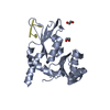
- PDB-7z3x: Crystal structure of FIR RRM1-2 Y115F mutant bound to FUSE ssDNA -

+
Open data


ID or keywords:

Loading...

-
Basic information

Entry
Database: PDB / ID: 7z3x
TitleCrystal structure of FIR RRM1-2 Y115F mutant bound to FUSE ssDNA
Components
  • DNA (5'-D(*TP*TP*T)-3')
  • DNA (5'-D(P*GP*TP*())-3')
  • Poly(U)-binding-splicing factor PUF60
KeywordsDNA BINDING PROTEIN / RNA recognition motif (RRM) / ssDNA
Function / homology
Function and homology information


alternative mRNA splicing, via spliceosome / mRNA splice site recognition / regulation of alternative mRNA splicing, via spliceosome / mRNA Splicing - Major Pathway / cell junction / cadherin binding / ribonucleoprotein complex / apoptotic process / DNA binding / RNA binding ...alternative mRNA splicing, via spliceosome / mRNA splice site recognition / regulation of alternative mRNA splicing, via spliceosome / mRNA Splicing - Major Pathway / cell junction / cadherin binding / ribonucleoprotein complex / apoptotic process / DNA binding / RNA binding / nucleoplasm / identical protein binding
Similarity search - Function
Poly-U binding splicing factor, PUF60-like / PUF60, RNA recognition motif 1 / PUF60, RNA recognition motif 2 / PUF60, RNA recognition motif 3 / RNA recognition motif domain, eukaryote / RNA recognition motif / RNA recognition motif / RNA recognition motif / Eukaryotic RNA Recognition Motif (RRM) profile. / RNA recognition motif domain ...Poly-U binding splicing factor, PUF60-like / PUF60, RNA recognition motif 1 / PUF60, RNA recognition motif 2 / PUF60, RNA recognition motif 3 / RNA recognition motif domain, eukaryote / RNA recognition motif / RNA recognition motif / RNA recognition motif / Eukaryotic RNA Recognition Motif (RRM) profile. / RNA recognition motif domain / RNA-binding domain superfamily / Nucleotide-binding alpha-beta plait domain superfamily
Similarity search - Domain/homology
DNA / Poly(U)-binding-splicing factor PUF60
Similarity search - Component
Biological speciesHomo sapiens (human)
MethodX-RAY DIFFRACTION / SYNCHROTRON / MOLECULAR REPLACEMENT / Resolution: 1.65 Å
AuthorsNi, X. / Chaikuad, A. / Knapp, S. / Structural Genomics Consortium (SGC)
Funding support1items
OrganizationGrant numberCountry
Not funded
CitationJournal: To Be Published
Title: Crystal structure of FIR RRM1-2 Y115F mutant bound to FUSE ssDNA
Authors: Ni, X. / Chaikuad, A. / Knapp, S.
History
DepositionMar 2, 2022Deposition site: PDBE / Processing site: PDBE
Revision 1.0Nov 9, 2022Provider: repository / Type: Initial release
Revision 1.1Nov 23, 2022Group: Derived calculations / Category: pdbx_struct_assembly / pdbx_struct_assembly_gen
Revision 2.0Jan 11, 2023Group: Advisory / Atomic model ...Advisory / Atomic model / Data collection / Database references / Derived calculations / Refinement description / Source and taxonomy / Structure summary
Category: atom_site / atom_site_anisotrop ...atom_site / atom_site_anisotrop / entity / entity_name_com / entity_src_gen / pdbx_contact_author / pdbx_initial_refinement_model / pdbx_nonpoly_scheme / pdbx_poly_seq_scheme / pdbx_refine_tls / pdbx_refine_tls_group / pdbx_struct_assembly / pdbx_struct_assembly_gen / pdbx_struct_sheet_hbond / pdbx_unobs_or_zero_occ_atoms / pdbx_unobs_or_zero_occ_residues / pdbx_validate_torsion / refine / refine_hist / refine_ls_restr / refine_ls_restr_ncs / refine_ls_shell / software / struct_conf / struct_mon_prot_cis / struct_ncs_dom / struct_ncs_dom_lim / struct_ref / struct_ref_seq / struct_ref_seq_dif / struct_sheet / struct_sheet_order / struct_sheet_range
Item: _entity.pdbx_description / _entity.pdbx_number_of_molecules ..._entity.pdbx_description / _entity.pdbx_number_of_molecules / _entity_name_com.name / _entity_src_gen.pdbx_gene_src_gene / _pdbx_poly_seq_scheme.auth_mon_id / _pdbx_poly_seq_scheme.auth_seq_num / _pdbx_poly_seq_scheme.pdb_mon_id / _pdbx_struct_sheet_hbond.range_1_auth_atom_id / _pdbx_struct_sheet_hbond.range_1_auth_comp_id / _pdbx_struct_sheet_hbond.range_1_auth_seq_id / _pdbx_struct_sheet_hbond.range_1_label_atom_id / _pdbx_struct_sheet_hbond.range_1_label_comp_id / _pdbx_struct_sheet_hbond.range_1_label_seq_id / _pdbx_struct_sheet_hbond.range_2_auth_atom_id / _pdbx_struct_sheet_hbond.range_2_auth_comp_id / _pdbx_struct_sheet_hbond.range_2_auth_seq_id / _pdbx_struct_sheet_hbond.range_2_label_atom_id / _pdbx_struct_sheet_hbond.range_2_label_comp_id / _pdbx_struct_sheet_hbond.range_2_label_seq_id / _pdbx_struct_sheet_hbond.range_id_1 / _pdbx_struct_sheet_hbond.range_id_2 / _pdbx_struct_sheet_hbond.sheet_id / _pdbx_validate_torsion.phi / _pdbx_validate_torsion.psi / _refine.B_iso_max / _refine.B_iso_mean / _refine.B_iso_min / _refine.ls_R_factor_R_free / _refine.ls_R_factor_R_work / _refine.ls_R_factor_obs / _refine.ls_number_reflns_R_work / _refine.pdbx_overall_phase_error / _refine_hist.cycle_id / _refine_hist.number_atoms_solvent / _refine_hist.number_atoms_total / _refine_hist.pdbx_B_iso_mean_ligand / _refine_hist.pdbx_B_iso_mean_solvent / _refine_hist.pdbx_number_atoms_protein / _refine_hist.pdbx_number_residues_total / _refine_ls_shell.R_factor_R_free / _refine_ls_shell.R_factor_R_free_error / _refine_ls_shell.R_factor_R_work / _refine_ls_shell.number_reflns_all / _refine_ls_shell.pdbx_total_number_of_bins_used / _software.version / _struct_conf.beg_auth_comp_id / _struct_conf.beg_auth_seq_id / _struct_conf.beg_label_comp_id / _struct_conf.beg_label_seq_id / _struct_conf.pdbx_PDB_helix_length / _struct_mon_prot_cis.pdbx_omega_angle / _struct_ref.db_code / _struct_ref.pdbx_align_begin / _struct_ref.pdbx_db_accession / _struct_ref.pdbx_seq_one_letter_code / _struct_ref_seq.db_align_beg / _struct_ref_seq.db_align_end / _struct_ref_seq.pdbx_auth_seq_align_beg / _struct_ref_seq.pdbx_db_accession / _struct_ref_seq.seq_align_beg / _struct_sheet_order.range_id_1 / _struct_sheet_order.range_id_2 / _struct_sheet_order.sheet_id
Description: Model completeness / Provider: author / Type: Coordinate replacement
Revision 2.1Feb 7, 2024Group: Data collection / Category: chem_comp_atom / chem_comp_bond

-
Structure visualization

Structure viewerMolecule:
MolmilJmol/JSmol

Downloads & links

-
Assembly

Deposited unit
A: Poly(U)-binding-splicing factor PUF60
B: Poly(U)-binding-splicing factor PUF60
D: DNA (5'-D(P*GP*TP*())-3')
E: DNA (5'-D(*TP*TP*T)-3')
hetero molecules


Theoretical massNumber of molelcules
Total (without water)45,6168
Polymers45,3684
Non-polymers2484
Water3,189177
1
A: Poly(U)-binding-splicing factor PUF60
D: DNA (5'-D(P*GP*TP*())-3')
hetero molecules


Theoretical massNumber of molelcules
Total (without water)22,8214
Polymers22,6972
Non-polymers1242
Water362
TypeNameSymmetry operationNumber
identity operation1_555x,y,z1
2
B: Poly(U)-binding-splicing factor PUF60
E: DNA (5'-D(*TP*TP*T)-3')
hetero molecules


Theoretical massNumber of molelcules
Total (without water)22,7964
Polymers22,6722
Non-polymers1242
Water181
TypeNameSymmetry operationNumber
identity operation1_555x,y,z1
Unit cell
Length a, b, c (Å)55.613, 70.386, 107.353
Angle α, β, γ (deg.)90.00, 90.00, 90.00
Int Tables number19
Space group name H-MP212121

-
Components

#1: Protein Poly(U)-binding-splicing factor PUF60 / 60 kDa poly(U)-binding-splicing factor / FUSE-binding protein-interacting repressor / FBP- ...60 kDa poly(U)-binding-splicing factor / FUSE-binding protein-interacting repressor / FBP-interacting repressor / Ro-binding protein 1 / RoBP1 / Siah-binding protein 1 / Siah-BP1


Mass: 21803.891 Da / Num. of mol.: 2
Source method: isolated from a genetically manipulated source
Source: (gene. exp.) Homo sapiens (human) / Gene: PUF60, FIR, ROBPI, SIAHBP1 / Production host: Escherichia coli BL21(DE3) (bacteria) / References: UniProt: Q9UHX1
#2: DNA chain DNA (5'-D(P*GP*TP*())-3')


Mass: 892.634 Da / Num. of mol.: 1 / Source method: obtained synthetically / Source: (synth.) Homo sapiens (human)
#3: DNA chain DNA (5'-D(*TP*TP*T)-3')


Mass: 867.621 Da / Num. of mol.: 1 / Source method: obtained synthetically / Source: (synth.) Homo sapiens (human)
#4: Chemical
ChemComp-EDO / 1,2-ETHANEDIOL / ETHYLENE GLYCOL / Ethylene glycol


Mass: 62.068 Da / Num. of mol.: 4 / Source method: obtained synthetically / Formula: C2H6O2
#5: Water ChemComp-HOH / water / Water


Mass: 18.015 Da / Num. of mol.: 177 / Source method: isolated from a natural source / Formula: H2O
Has ligand of interestN

-
Experimental details

-
Experiment

ExperimentMethod: X-RAY DIFFRACTION / Number of used crystals: 1

-
Sample preparation

CrystalDensity Matthews: 2.33 Å3/Da / Density % sol: 47.26 %
Crystal growTemperature: 293 K / Method: vapor diffusion, sitting drop / pH: 5.5 / Details: 25% PEG3350 0.1M Bis-Tris, pH5.5

-
Data collection

DiffractionMean temperature: 100 K / Serial crystal experiment: N
Diffraction sourceSource: SYNCHROTRON / Site: SLS / Beamline: X06SA / Wavelength: 0.999998 Å
DetectorType: DECTRIS EIGER X 16M / Detector: PIXEL / Date: Dec 15, 2021
RadiationProtocol: SINGLE WAVELENGTH / Monochromatic (M) / Laue (L): M / Scattering type: x-ray
Radiation wavelengthWavelength: 0.999998 Å / Relative weight: 1
ReflectionResolution: 1.65→49.38 Å / Num. obs: 50994 / % possible obs: 99.2 % / Redundancy: 4.3 % / Biso Wilson estimate: 27.18 Å2 / CC1/2: 0.999 / Net I/σ(I): 16.3
Reflection shellResolution: 1.65→1.68 Å / Num. unique obs: 2538 / CC1/2: 0.833

-
Processing

Software
NameVersionClassification
PHENIX1.2-4459refinement
Aimlessdata scaling
PDB_EXTRACT3.27data extraction
XDSdata reduction
PHASERphasing
RefinementMethod to determine structure: MOLECULAR REPLACEMENT
Starting model: 2QFJ
Resolution: 1.65→49.38 Å / SU ML: 0.24 / Cross valid method: THROUGHOUT / σ(F): 1.37 / Phase error: 22.39 / Stereochemistry target values: ML
RfactorNum. reflection% reflection
Rfree0.2144 2554 5.01 %
Rwork0.2038 --
obs0.2044 50947 98.99 %
Solvent computationShrinkage radii: 0.9 Å / VDW probe radii: 1.1 Å / Solvent model: FLAT BULK SOLVENT MODEL
Refinement stepCycle: LAST / Resolution: 1.65→49.38 Å
ProteinNucleic acidLigandSolventTotal
Num. atoms2875 87 16 177 3155
Refine LS restraints
Refine-IDTypeDev idealNumber
X-RAY DIFFRACTIONf_bond_d0.007
X-RAY DIFFRACTIONf_angle_d1.041
X-RAY DIFFRACTIONf_dihedral_angle_d9.207476
X-RAY DIFFRACTIONf_chiral_restr0.059466
X-RAY DIFFRACTIONf_plane_restr0.008546
LS refinement shell
Resolution (Å)Rfactor RfreeNum. reflection RfreeRfactor RworkNum. reflection RworkRefine-ID% reflection obs (%)
1.65-1.680.38711340.33652683X-RAY DIFFRACTION100
1.68-1.720.35451490.30042653X-RAY DIFFRACTION100
1.72-1.750.30661450.26752683X-RAY DIFFRACTION100
1.75-1.790.30511350.2472684X-RAY DIFFRACTION100
1.79-1.840.24931560.23062606X-RAY DIFFRACTION99
1.84-1.890.24661190.22422663X-RAY DIFFRACTION98
1.89-1.940.26661540.22842670X-RAY DIFFRACTION100
1.94-2.010.26191610.24182658X-RAY DIFFRACTION100
2.01-2.080.27291440.23122695X-RAY DIFFRACTION100
2.08-2.160.20911400.20712664X-RAY DIFFRACTION100
2.16-2.260.22271350.1972703X-RAY DIFFRACTION100
2.26-2.380.2441320.20812717X-RAY DIFFRACTION100
2.38-2.530.2491350.21242711X-RAY DIFFRACTION99
2.53-2.720.24451410.2132606X-RAY DIFFRACTION96
2.72-30.23211180.20852748X-RAY DIFFRACTION99
3-3.430.22481540.20752725X-RAY DIFFRACTION99
3.43-4.320.17111530.16862748X-RAY DIFFRACTION99
4.32-49.380.15631490.18632776X-RAY DIFFRACTION95
Refinement TLS params.

Method: refined / Refine-ID: X-RAY DIFFRACTION

IDL112)L122)L132)L222)L232)L332)S11 (Å °)S12 (Å °)S13 (Å °)S21 (Å °)S22 (Å °)S23 (Å °)S31 (Å °)S32 (Å °)S33 (Å °)T112)T122)T132)T222)T232)T332)Origin x (Å)Origin y (Å)Origin z (Å)
15.54781.9078-3.60846.3879-3.48528.68-0.05160.02520.3395-0.53420.1128-0.3758-0.12460.3285-0.08980.3144-0.0447-0.0060.1563-0.00390.271-2.97575.48119.7561
21.93540.31811.11583.17250.3612.8696-0.0274-0.46880.24810.38010.05710.1355-0.3794-0.6445-0.07830.28620.08540.01950.3009-0.03010.208-11.8553-1.132142.8833
35.51820.3283-2.10612.4399-1.78316.3192-0.0913-0.10350.17390.00850.0379-0.0969-0.1623-0.13790.08720.1790.0287-0.01010.1683-0.00610.1497-7.0484-6.006732.0902
41.72790.8073-0.61383.5099-3.07029.2942-0.0684-0.19110.04470.2750.19840.1942-0.161-0.9031-0.16190.16550.02170.01090.24120.00450.189-15.7938-5.23132.8674
59.3021-6.3032-0.4188.2619-0.06196.08550.54130.8422-0.0231-1.1809-0.33010.16050.12450.2089-0.15950.31380.001-0.00980.20620.00520.1513-5.0943-5.915414.3087
63.2737-0.5925-0.84365.6683-0.34295.03340.0171-0.02190.18520.0001-0.1261-0.5640.04970.57910.06490.13380.0465-0.00270.17980.02280.2236.8703-15.436229.0899
74.3775-1.65571.58956.4274-0.93483.5269-0.09920.40230.25920.06340.0716-0.5621-0.28570.15820.26710.19210.01090.02250.1866-0.00040.21870.3825-9.624523.0557
85.5136-0.0768-0.27030.69780.89691.17040.10260.221-0.413-0.10860.36490.91110.0359-0.8641-0.22070.321-0.0351-0.09720.62970.27940.5666-37.2317-1.18228.2369
91.8587-1.13510.97864.7184-3.52775.0305-0.0037-0.1361-0.06620.01260.23690.2736-0.1268-0.2298-0.15740.19490.0111-0.01050.17780.01370.185-21.37354.77947.1351
100.26341.1062-0.19864.8436-1.17173.46530.0711-0.432-0.07740.2126-0.04190.9283-0.4996-0.0535-0.07020.30810.02480.09850.62570.20320.7326-40.6816-4.396919.0608
114.3503-0.0179-0.7753.0478-1.13771.1127-0.535-1.1073-1.28550.07810.65620.3310.4453-0.18270.05380.31010.06660.02260.54150.27640.505-22.6283-15.756719.2307
122.79130.0796-1.92853.0363-2.64323.57410.70620.00620.6044-0.2494-0.16891.114-1.0123-1.8271-0.39970.54060.1832-0.00470.74850.06590.4526-22.37911.553135.6201
136.77384.3704-2.84857.41711.35196.2956-0.51260.65380.697-1.03150.8931.3542-0.3984-0.8975-0.47660.50830.1268-0.07440.6470.19680.6032-30.893811.86995.2408
Refinement TLS group
IDRefine-IDRefine TLS-IDSelection details
1X-RAY DIFFRACTION1chain 'A' and (resid 98 through 111 )
2X-RAY DIFFRACTION2chain 'A' and (resid 112 through 155 )
3X-RAY DIFFRACTION3chain 'A' and (resid 156 through 173 )
4X-RAY DIFFRACTION4chain 'A' and (resid 174 through 196 )
5X-RAY DIFFRACTION5chain 'A' and (resid 197 through 207 )
6X-RAY DIFFRACTION6chain 'A' and (resid 208 through 280 )
7X-RAY DIFFRACTION7chain 'A' and (resid 281 through 291 )
8X-RAY DIFFRACTION8chain 'B' and (resid 99 through 117 )
9X-RAY DIFFRACTION9chain 'B' and (resid 118 through 191 )
10X-RAY DIFFRACTION10chain 'B' and (resid 192 through 208 )
11X-RAY DIFFRACTION11chain 'B' and (resid 209 through 289 )
12X-RAY DIFFRACTION12chain 'D' and (resid 1 through 3 )
13X-RAY DIFFRACTION13chain 'E' and (resid 0 through 2 )

+
About Yorodumi

-
News

-
Feb 9, 2022. New format data for meta-information of EMDB entries

New format data for meta-information of EMDB entries

  • Version 3 of the EMDB header file is now the official format.
  • The previous official version 1.9 will be removed from the archive.

Related info.:EMDB header

External links:wwPDB to switch to version 3 of the EMDB data model

-
Aug 12, 2020. Covid-19 info

Covid-19 info

URL: https://pdbj.org/emnavi/covid19.php

New page: Covid-19 featured information page in EM Navigator.

Related info.:Covid-19 info / Mar 5, 2020. Novel coronavirus structure data

+
Mar 5, 2020. Novel coronavirus structure data

Novel coronavirus structure data

Related info.:Yorodumi Speices / Aug 12, 2020. Covid-19 info

External links:COVID-19 featured content - PDBj / Molecule of the Month (242):Coronavirus Proteases

+
Jan 31, 2019. EMDB accession codes are about to change! (news from PDBe EMDB page)

EMDB accession codes are about to change! (news from PDBe EMDB page)

  • The allocation of 4 digits for EMDB accession codes will soon come to an end. Whilst these codes will remain in use, new EMDB accession codes will include an additional digit and will expand incrementally as the available range of codes is exhausted. The current 4-digit format prefixed with “EMD-” (i.e. EMD-XXXX) will advance to a 5-digit format (i.e. EMD-XXXXX), and so on. It is currently estimated that the 4-digit codes will be depleted around Spring 2019, at which point the 5-digit format will come into force.
  • The EM Navigator/Yorodumi systems omit the EMD- prefix.

Related info.:Q: What is EMD? / ID/Accession-code notation in Yorodumi/EM Navigator

External links:EMDB Accession Codes are Changing Soon! / Contact to PDBj

+
Jul 12, 2017. Major update of PDB

Major update of PDB

  • wwPDB released updated PDB data conforming to the new PDBx/mmCIF dictionary.
  • This is a major update changing the version number from 4 to 5, and with Remediation, in which all the entries are updated.
  • In this update, many items about electron microscopy experimental information are reorganized (e.g. em_software).
  • Now, EM Navigator and Yorodumi are based on the updated data.

External links:wwPDB Remediation / Enriched Model Files Conforming to OneDep Data Standards Now Available in the PDB FTP Archive

-
Yorodumi

Thousand views of thousand structures

  • Yorodumi is a browser for structure data from EMDB, PDB, SASBDB, etc.
  • This page is also the successor to EM Navigator detail page, and also detail information page/front-end page for Omokage search.
  • The word "yorodu" (or yorozu) is an old Japanese word meaning "ten thousand". "mi" (miru) is to see.

Related info.:EMDB / PDB / SASBDB / Comparison of 3 databanks / Yorodumi Search / Aug 31, 2016. New EM Navigator & Yorodumi / Yorodumi Papers / Jmol/JSmol / Function and homology information / Changes in new EM Navigator and Yorodumi

Read more