 Movie
Movie Controller

[English] 日本語
 Yorodumi
- PDB-7z3b: Crystal structure of the cupredoxin AcoP from Acidithiobacillus f... -
+ Open data

- Basic information
| Entry | Database: PDB / ID: 7z3b | ||||||
|---|---|---|---|---|---|---|---|
| Title | Crystal structure of the cupredoxin AcoP from Acidithiobacillus ferrooxidans, reduced form | ||||||
 Components Components | AcoP | ||||||
 Keywords Keywords | METAL BINDING PROTEIN / cupredoxin / copper-binding | ||||||
| Function / homology | Cupredoxin / metal ion binding / ACETATE ION / COPPER (I) ION / EfeO-type cupredoxin-like domain-containing protein Function and homology information Function and homology information | ||||||
| Biological species |  Acidithiobacillus ferrooxidans (bacteria) Acidithiobacillus ferrooxidans (bacteria) | ||||||
| Method |  X-RAY DIFFRACTION / SYNCHROTRON / SAD / Resolution: 1.65 Å X-RAY DIFFRACTION / SYNCHROTRON / SAD / Resolution: 1.65 Å | ||||||
 Authors Authors | Leone, P. / Sciara, G. / Ilbert, M. | ||||||
| Funding support |  France, 1items France, 1items
| ||||||
 Citation Citation | Journal: Dalton Trans / Year: 2024 Title: Beyond the coupled distortion model: structural analysis of the single domain cupredoxin AcoP, a green mononuclear copper centre with original features. Authors: Roger, M. / Leone, P. / Blackburn, N.J. / Horrell, S. / Chicano, T.M. / Biaso, F. / Giudici-Orticoni, M.T. / Abriata, L.A. / Hura, G.L. / Hough, M.A. / Sciara, G. / Ilbert, M. | ||||||
| History |
|
- Structure visualization
| Structure viewer | Molecule: MolmilJmol/JSmol |
|---|
- Downloads & links
-Download
| PDBx/mmCIF format | 7z3b.cif.gz | 131.7 KB | Display | PDBx/mmCIF format |
|---|---|---|---|---|
| PDB format | pdb7z3b.ent.gz | 101.1 KB | Display | PDB format |
| PDBx/mmJSON format | 7z3b.json.gz | Tree view | PDBx/mmJSON format | |
| Others |  Other downloads Other downloads |
-Validation report
| Summary document | 7z3b_validation.pdf.gz | 1.2 MB | Display | wwPDB validaton report |
|---|---|---|---|---|
| Full document | 7z3b_full_validation.pdf.gz | 1.2 MB | Display | |
| Data in XML | 7z3b_validation.xml.gz | 14.7 KB | Display | |
| Data in CIF | 7z3b_validation.cif.gz | 20.7 KB | Display | |
| Arichive directory | https://data.pdbj.org/pub/pdb/validation_reports/z3/7z3bftp://data.pdbj.org/pub/pdb/validation_reports/z3/7z3b | HTTPS FTP |
-Related structure data


-Links
 PDBj
PDBj- Assembly
| Deposited unit | 
| ||||||||||||||||||
|---|---|---|---|---|---|---|---|---|---|---|---|---|---|---|---|---|---|---|---|
| 1 | 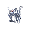
| ||||||||||||||||||
| 2 | 
| ||||||||||||||||||
| Unit cell |
| ||||||||||||||||||
| Noncrystallographic symmetry (NCS) | NCS domain:
NCS domain segments: Component-ID: _ / Ens-ID: 1 / Beg auth comp-ID: GLY / Beg label comp-ID: GLY / End auth comp-ID: GLN / End label comp-ID: GLN / Refine code: _ / Auth seq-ID: 43 - 181 / Label seq-ID: 31 - 169
|
-Components
| #1: Protein | Mass: 19105.320 Da / Num. of mol.: 2 Source method: isolated from a genetically manipulated source Source: (gene. exp.) Acidithiobacillus ferrooxidans (bacteria)Gene: DN052_11965 / Production host: #2: Chemical |   Mass: 63.546 Da / Num. of mol.: 2 / Source method: obtained synthetically / Formula: Cu / Feature type: SUBJECT OF INVESTIGATION Mass: 63.546 Da / Num. of mol.: 2 / Source method: obtained synthetically / Formula: Cu / Feature type: SUBJECT OF INVESTIGATION#3: Chemical |   Mass: 92.094 Da / Num. of mol.: 3 / Source method: obtained synthetically / Formula: C3H8O3 Mass: 92.094 Da / Num. of mol.: 3 / Source method: obtained synthetically / Formula: C3H8O3#4: Chemical |   Mass: 59.044 Da / Num. of mol.: 2 / Source method: obtained synthetically / Formula: C2H3O2 Mass: 59.044 Da / Num. of mol.: 2 / Source method: obtained synthetically / Formula: C2H3O2#5: Water | ChemComp-HOH / |  Mass: 18.015 Da / Num. of mol.: 173 / Source method: isolated from a natural source / Formula: H2O Mass: 18.015 Da / Num. of mol.: 173 / Source method: isolated from a natural source / Formula: H2OHas ligand of interest | Y | |
|---|
-Experimental details
-Experiment
| Experiment | Method: X-RAY DIFFRACTION / Number of used crystals: 1 |
|---|
- Sample preparation
| Crystal | Density Matthews: 1.96 Å3/Da / Density % sol: 37.38 % |
|---|---|
| Crystal grow | Temperature: 293 K / Method: vapor diffusion, sitting drop Details: 100mM potassium acetate, 10mM potassium chloride, 50mM MES, 50mM Tris, 50mM Hepes, 34-44% PEG3000 PH range: 6.0-8.0 |
-Data collection
| Diffraction | Mean temperature: 100 K / Serial crystal experiment: N |
|---|---|
| Diffraction source | Source: SYNCHROTRON / Site: SOLEIL / Beamline: PROXIMA 1 / Wavelength: 1.07137 Å |
| Detector | Type: PSI JUNGFRAU 4M / Detector: PIXEL / Date: Jun 2, 2015 |
| Radiation | Protocol: SINGLE WAVELENGTH / Monochromatic (M) / Laue (L): M / Scattering type: x-ray |
| Radiation wavelength | Wavelength: 1.07137 Å / Relative weight: 1 |
| Reflection | Resolution: 1.65→50 Å / Num. obs: 37412 / % possible obs: 99.9 % / Redundancy: 6.9 % / CC1/2: 0.999 / Net I/σ(I): 15.4 |
| Reflection shell | Resolution: 1.65→1.74 Å / Num. unique obs: 5387 / CC1/2: 0.638 |
- Processing
| Software |
| |||||||||||||||||||||||||||||||||||||||||||||||||||||||||||||||||||||||||||
|---|---|---|---|---|---|---|---|---|---|---|---|---|---|---|---|---|---|---|---|---|---|---|---|---|---|---|---|---|---|---|---|---|---|---|---|---|---|---|---|---|---|---|---|---|---|---|---|---|---|---|---|---|---|---|---|---|---|---|---|---|---|---|---|---|---|---|---|---|---|---|---|---|---|---|---|---|
| Refinement | Method to determine structure: SAD / Resolution: 1.65→44.63 Å / Cor.coef. Fo:Fc: 0.973 / Cor.coef. Fo:Fc free: 0.96 / SU B: 3.7 / SU ML: 0.06 / Cross valid method: THROUGHOUT / σ(F): 0 / ESU R: 0.082 / ESU R Free: 0.084 / Stereochemistry target values: MAXIMUM LIKELIHOOD Details: U VALUES : WITH TLS ADDED HYDROGENS HAVE BEEN ADDED IN THE RIDING POSITIONS
| |||||||||||||||||||||||||||||||||||||||||||||||||||||||||||||||||||||||||||
| Solvent computation | Ion probe radii: 0.8 Å / Shrinkage radii: 0.8 Å / VDW probe radii: 1.2 Å / Solvent model: MASK | |||||||||||||||||||||||||||||||||||||||||||||||||||||||||||||||||||||||||||
| Displacement parameters | Biso max: 107.45 Å2 / Biso mean: 33.374 Å2 / Biso min: 17.31 Å2
| |||||||||||||||||||||||||||||||||||||||||||||||||||||||||||||||||||||||||||
| Refinement step | Cycle: final / Resolution: 1.65→44.63 Å
| |||||||||||||||||||||||||||||||||||||||||||||||||||||||||||||||||||||||||||
| Refine LS restraints |
| |||||||||||||||||||||||||||||||||||||||||||||||||||||||||||||||||||||||||||
| Refine LS restraints NCS | Ens-ID: 1 / Number: 4165 / Refine-ID: X-RAY DIFFRACTION / Type: interatomic distance / Rms dev position: 0.13 Å / Weight position: 0.05
| |||||||||||||||||||||||||||||||||||||||||||||||||||||||||||||||||||||||||||
| LS refinement shell | Resolution: 1.65→1.693 Å / Rfactor Rfree error: 0 / Total num. of bins used: 20
| |||||||||||||||||||||||||||||||||||||||||||||||||||||||||||||||||||||||||||
| Refinement TLS params. | Method: refined / Refine-ID: X-RAY DIFFRACTION
| |||||||||||||||||||||||||||||||||||||||||||||||||||||||||||||||||||||||||||
| Refinement TLS group |
|
