 Movie
Movie Controller


+ Open data

- Basic information
| Entry | Database: PDB / ID: 7z0i | ||||||
|---|---|---|---|---|---|---|---|
| Title | human PEX13 SH3 domain | ||||||
 Components Components | Peroxisomal membrane protein PEX13 | ||||||
 Keywords Keywords | PROTEIN TRANSPORT | ||||||
| Function / homology |  Function and homology information Function and homology informationmicrotubule-based peroxisome localization / protein import into peroxisome matrix, translocation / peroxisomal importomer complex / fatty acid alpha-oxidation / protein import into peroxisome matrix, docking / Class I peroxisomal membrane protein import / suckling behavior / peroxisomal membrane / cerebral cortex cell migration / transmembrane protein transporter activity ...microtubule-based peroxisome localization / protein import into peroxisome matrix, translocation / peroxisomal importomer complex / fatty acid alpha-oxidation / protein import into peroxisome matrix, docking / Class I peroxisomal membrane protein import / suckling behavior / peroxisomal membrane / cerebral cortex cell migration / transmembrane protein transporter activity / cellular response to reactive oxygen species / Peroxisomal protein import / locomotory behavior / neuron migration / peroxisome / E3 ubiquitin ligases ubiquitinate target proteins / membrane / cytosol Similarity search - Function | ||||||
| Biological species |  Homo sapiens (human) Homo sapiens (human) | ||||||
| Method |  X-RAY DIFFRACTION / SYNCHROTRON / MOLECULAR REPLACEMENT / Resolution: 1.8 Å X-RAY DIFFRACTION / SYNCHROTRON / MOLECULAR REPLACEMENT / Resolution: 1.8 Å | ||||||
 Authors Authors | Gaussmann, S. / Zak, K. / Sattler, M. | ||||||
| Funding support |  Germany, 1items Germany, 1items
| ||||||
 Citation Citation | Journal: Nat Commun / Year: 2024 Title: Modulation of peroxisomal import by the PEX13 SH3 domain and a proximal FxxxF binding motif. Authors: Gaussmann, S. / Peschel, R. / Ott, J. / Zak, K.M. / Sastre, J. / Delhommel, F. / Popowicz, G.M. / Boekhoven, J. / Schliebs, W. / Erdmann, R. / Sattler, M. #1: Journal: Biorxiv / Year: 2022Title: Intramolecular autoinhibition of human PEX13 modulates peroxisomal import Authors: Gaussmann, S. / Ott, J. / Zak, K.M. / Delhommel, F. / Popowicz, G.M. / Schliebs, W. / Erdmann, R. / Sattler, M. | ||||||
| History |
|
- Structure visualization
| Structure viewer | Molecule: MolmilJmol/JSmol |
|---|
- Downloads & links
-Download
| PDBx/mmCIF format | 7z0i.cif.gz | 47 KB | Display | PDBx/mmCIF format |
|---|---|---|---|---|
| PDB format | pdb7z0i.ent.gz | 31.1 KB | Display | PDB format |
| PDBx/mmJSON format | 7z0i.json.gz | Tree view | PDBx/mmJSON format | |
| Others |  Other downloads Other downloads |
-Validation report
| Arichive directory | https://data.pdbj.org/pub/pdb/validation_reports/z0/7z0iftp://data.pdbj.org/pub/pdb/validation_reports/z0/7z0i | HTTPS FTP |
|---|
-Related structure data
| Related structure data | 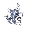 7z0jC  7z0kC 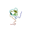 1wxuS S: Starting model for refinement C: citing same article ( |
|---|---|
| Similar structure data |
-Links
 PDBj
PDBj
- Assembly
| Deposited unit | 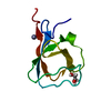
| ||||||||
|---|---|---|---|---|---|---|---|---|---|
| 1 |
| ||||||||
| Unit cell |
| ||||||||
| Components on special symmetry positions |
|
-Components
| #1: Protein | Mass: 9461.601 Da / Num. of mol.: 1 Source method: isolated from a genetically manipulated source Source: (gene. exp.) Homo sapiens (human) / Gene: PEX13 / Production host:  |
|---|---|
| #2: Chemical | ChemComp-ZN /   Mass: 65.409 Da / Num. of mol.: 1 / Source method: obtained synthetically / Formula: Zn Mass: 65.409 Da / Num. of mol.: 1 / Source method: obtained synthetically / Formula: Zn |
| #3: Chemical | ChemComp-EDO /   Mass: 62.068 Da / Num. of mol.: 1 / Source method: isolated from a natural source / Formula: C2H6O2 Mass: 62.068 Da / Num. of mol.: 1 / Source method: isolated from a natural source / Formula: C2H6O2 |
| #4: Water | ChemComp-HOH /  Mass: 18.015 Da / Num. of mol.: 51 / Source method: isolated from a natural source / Formula: H2O Mass: 18.015 Da / Num. of mol.: 51 / Source method: isolated from a natural source / Formula: H2O |
| Has ligand of interest | N |
| Has protein modification | N |
-Experimental details
-Experiment
| Experiment | Method: X-RAY DIFFRACTION / Number of used crystals: 1 |
|---|
- Sample preparation
| Crystal | Density Matthews: 2.36 Å3/Da / Density % sol: 48 % |
|---|---|
| Crystal grow | Temperature: 293 K / Method: vapor diffusion, sitting drop / pH: 5 Details: 0.01M Zinc chloride, 0.1M Sodium acetate, 20% w/v PEG 6000 |
-Data collection
| Diffraction | Mean temperature: 100 K / Serial crystal experiment: N | ||||||||||||||||||||||||
|---|---|---|---|---|---|---|---|---|---|---|---|---|---|---|---|---|---|---|---|---|---|---|---|---|---|
| Diffraction source | Source: SYNCHROTRON / Site: SLS  / Beamline: X06DA / Wavelength: 0.999995 Å / Beamline: X06DA / Wavelength: 0.999995 Å | ||||||||||||||||||||||||
| Detector | Type: DECTRIS PILATUS 2M-F / Detector: PIXEL / Date: Feb 8, 2020 | ||||||||||||||||||||||||
| Radiation | Protocol: SINGLE WAVELENGTH / Monochromatic (M) / Laue (L): M / Scattering type: x-ray | ||||||||||||||||||||||||
| Radiation wavelength | Wavelength: 0.999995 Å / Relative weight: 1 | ||||||||||||||||||||||||
| Reflection | Resolution: 1.8→43.39 Å / Num. obs: 7660 / % possible obs: 99.9 % / Redundancy: 13.6 % / CC1/2: 1 / Rmerge(I) obs: 0.082 / Rpim(I) all: 0.034 / Rrim(I) all: 0.089 / Net I/σ(I): 19.7 | ||||||||||||||||||||||||
| Reflection shell | Diffraction-ID: 1
|
- Processing
| Software |
| |||||||||||||||||||||||||||||||||||||||||||||||||||||||||||||||||||||||||||||||||||||||||||||||||||||||||||||||||||||||||||||||||||||||||||||||||||||||||||
|---|---|---|---|---|---|---|---|---|---|---|---|---|---|---|---|---|---|---|---|---|---|---|---|---|---|---|---|---|---|---|---|---|---|---|---|---|---|---|---|---|---|---|---|---|---|---|---|---|---|---|---|---|---|---|---|---|---|---|---|---|---|---|---|---|---|---|---|---|---|---|---|---|---|---|---|---|---|---|---|---|---|---|---|---|---|---|---|---|---|---|---|---|---|---|---|---|---|---|---|---|---|---|---|---|---|---|---|---|---|---|---|---|---|---|---|---|---|---|---|---|---|---|---|---|---|---|---|---|---|---|---|---|---|---|---|---|---|---|---|---|---|---|---|---|---|---|---|---|---|---|---|---|---|---|---|---|
| Refinement | Method to determine structure: MOLECULAR REPLACEMENT Starting model: 1wxu Resolution: 1.8→39.238 Å / Cor.coef. Fo:Fc: 0.967 / Cor.coef. Fo:Fc free: 0.946 / WRfactor Rfree: 0.219 / WRfactor Rwork: 0.172 / SU B: 3.501 / SU ML: 0.105 / Average fsc free: 0.8961 / Average fsc work: 0.9082 / Cross valid method: THROUGHOUT / ESU R: 0.128 / ESU R Free: 0.128 Details: Hydrogens have been added in their riding positions
| |||||||||||||||||||||||||||||||||||||||||||||||||||||||||||||||||||||||||||||||||||||||||||||||||||||||||||||||||||||||||||||||||||||||||||||||||||||||||||
| Solvent computation | Ion probe radii: 0.8 Å / Shrinkage radii: 0.8 Å / VDW probe radii: 1.2 Å / Solvent model: MASK BULK SOLVENT | |||||||||||||||||||||||||||||||||||||||||||||||||||||||||||||||||||||||||||||||||||||||||||||||||||||||||||||||||||||||||||||||||||||||||||||||||||||||||||
| Displacement parameters | Biso mean: 34.564 Å2
| |||||||||||||||||||||||||||||||||||||||||||||||||||||||||||||||||||||||||||||||||||||||||||||||||||||||||||||||||||||||||||||||||||||||||||||||||||||||||||
| Refinement step | Cycle: LAST / Resolution: 1.8→39.238 Å
| |||||||||||||||||||||||||||||||||||||||||||||||||||||||||||||||||||||||||||||||||||||||||||||||||||||||||||||||||||||||||||||||||||||||||||||||||||||||||||
| Refine LS restraints |
| |||||||||||||||||||||||||||||||||||||||||||||||||||||||||||||||||||||||||||||||||||||||||||||||||||||||||||||||||||||||||||||||||||||||||||||||||||||||||||
| LS refinement shell |
|
