 Movie
Movie Controller

[English] 日本語
 Yorodumi
- PDB-7yua: Structural Insight into a Metal-Dependent Mutase MtdL Revealing a... -
+ Open data

- Basic information
| Entry | Database: PDB / ID: 7yua | ||||||
|---|---|---|---|---|---|---|---|
| Title | Structural Insight into a Metal-Dependent Mutase MtdL Revealing an Arginine Residue Covalently Mediated Interconversion between Nucleotide-Based furanose and pyranose | ||||||
 Components Components | Transglycosylse | ||||||
 Keywords Keywords | BIOSYNTHETIC PROTEIN / Metal-Dependent Mutase | ||||||
| Function / homology | STELLO-like / Reversibly glycosylated polypeptide family / Reversibly glycosylated polypeptide / metal ion binding / GUANOSINE-5'-DIPHOSPHATE / Transglycosylse Function and homology information Function and homology information | ||||||
| Biological species |  Marinactinospora thermotolerans (bacteria) Marinactinospora thermotolerans (bacteria) | ||||||
| Method |  X-RAY DIFFRACTION / SYNCHROTRON / MOLECULAR REPLACEMENT / Resolution: 2.5 Å X-RAY DIFFRACTION / SYNCHROTRON / MOLECULAR REPLACEMENT / Resolution: 2.5 Å | ||||||
 Authors Authors | Chi, C.B. / Ma, M. | ||||||
| Funding support | 1items
| ||||||
 Citation Citation | Journal: Acs Catalysis / Year: 2023 Title: Structural Insight into a Metal-Dependent Mutase Revealing an Arginine Residue-Covalently Mediated Interconversion between Nucleotide-Based Pyranose and Furanose. Authors: Chi, C.B. / Xu, R. / Chen, Q.Q. / Zhang, X.H. / Shi, X.M. / Jin, H.W. / Yin, F. / Jia, H.L. / Zhang, L.R. / Yang, D.H. / Ju, J.H. / Li, Q.L. / Ma, M. | ||||||
| History |
|
- Structure visualization
| Structure viewer | Molecule: MolmilJmol/JSmol |
|---|
- Downloads & links
-Download
| PDBx/mmCIF format | 7yua.cif.gz | 313 KB | Display | PDBx/mmCIF format |
|---|---|---|---|---|
| PDB format | pdb7yua.ent.gz | 251.3 KB | Display | PDB format |
| PDBx/mmJSON format | 7yua.json.gz | Tree view | PDBx/mmJSON format | |
| Others |  Other downloads Other downloads |
-Validation report
| Summary document | 7yua_validation.pdf.gz | 2.1 MB | Display | wwPDB validaton report |
|---|---|---|---|---|
| Full document | 7yua_full_validation.pdf.gz | 2.2 MB | Display | |
| Data in XML | 7yua_validation.xml.gz | 64.1 KB | Display | |
| Data in CIF | 7yua_validation.cif.gz | 88.2 KB | Display | |
| Arichive directory | https://data.pdbj.org/pub/pdb/validation_reports/yu/7yuaftp://data.pdbj.org/pub/pdb/validation_reports/yu/7yua | HTTPS FTP |
-Related structure data
| Related structure data | 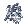 7yv0C  7yu9 S: Starting model for refinement C: citing same article ( |
|---|---|
| Similar structure data |
-Links
 PDBj
PDBj- Assembly
| Deposited unit | 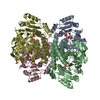
| ||||||||
|---|---|---|---|---|---|---|---|---|---|
| 1 |
| ||||||||
| Unit cell |
|
-Components
| #1: Protein | Mass: 41653.750 Da / Num. of mol.: 4 Source method: isolated from a genetically manipulated source Source: (gene. exp.) Marinactinospora thermotolerans (bacteria)Production host: References: UniProt: G8HX37 #2: Chemical | ChemComp-GDP /   Type: RNA linking / Mass: 443.201 Da / Num. of mol.: 4 / Source method: isolated from a natural source / Formula: C10H15N5O11P2 / Comment: GDP, energy-carrying molecule*YM Type: RNA linking / Mass: 443.201 Da / Num. of mol.: 4 / Source method: isolated from a natural source / Formula: C10H15N5O11P2 / Comment: GDP, energy-carrying molecule*YM#3: Chemical | ChemComp-MG /   Mass: 24.305 Da / Num. of mol.: 11 / Source method: obtained synthetically / Formula: Mg Mass: 24.305 Da / Num. of mol.: 11 / Source method: obtained synthetically / Formula: Mg#4: Chemical |   Mass: 96.063 Da / Num. of mol.: 2 / Source method: obtained synthetically / Formula: SO4 / Feature type: SUBJECT OF INVESTIGATION Mass: 96.063 Da / Num. of mol.: 2 / Source method: obtained synthetically / Formula: SO4 / Feature type: SUBJECT OF INVESTIGATION#5: Water | ChemComp-HOH / |  Mass: 18.015 Da / Num. of mol.: 569 / Source method: isolated from a natural source / Formula: H2O Mass: 18.015 Da / Num. of mol.: 569 / Source method: isolated from a natural source / Formula: H2OHas ligand of interest | Y | Has protein modification | Y | |
|---|
-Experimental details
-Experiment
| Experiment | Method: X-RAY DIFFRACTION / Number of used crystals: 1 |
|---|
- Sample preparation
| Crystal | Density Matthews: 2.6 Å3/Da / Density % sol: 52.65 % |
|---|---|
| Crystal grow | Temperature: 291 K / Method: vapor diffusion, hanging drop / Details: containing 18 % PEG 3350, 0.2 M Potassium Formate |
-Data collection
| Diffraction | Mean temperature: 100 K / Serial crystal experiment: N |
|---|---|
| Diffraction source | Source: SYNCHROTRON / Site: NFPSS  / Beamline: BL18U / Wavelength: 0.979 Å / Beamline: BL18U / Wavelength: 0.979 Å |
| Detector | Type: DECTRIS PILATUS3 6M / Detector: PIXEL / Date: Oct 17, 2019 |
| Radiation | Protocol: SINGLE WAVELENGTH / Monochromatic (M) / Laue (L): M / Scattering type: x-ray |
| Radiation wavelength | Wavelength: 0.979 Å / Relative weight: 1 |
| Reflection | Resolution: 2.5→49.91 Å / Num. obs: 58110 / % possible obs: 100 % / Redundancy: 2 % / Rmerge(I) obs: 0.085 / Net I/σ(I): 6.8 |
| Reflection shell | Resolution: 2.5→2.57 Å / Rmerge(I) obs: 0.412 / Num. unique obs: 4540 |
- Processing
| Software |
| ||||||||||||||||||||||||||||||||||||||||||||||||||||||||||||||||||||||||||||||||||||||||||||||||||||||||||||||||||||||||||||||||||||
|---|---|---|---|---|---|---|---|---|---|---|---|---|---|---|---|---|---|---|---|---|---|---|---|---|---|---|---|---|---|---|---|---|---|---|---|---|---|---|---|---|---|---|---|---|---|---|---|---|---|---|---|---|---|---|---|---|---|---|---|---|---|---|---|---|---|---|---|---|---|---|---|---|---|---|---|---|---|---|---|---|---|---|---|---|---|---|---|---|---|---|---|---|---|---|---|---|---|---|---|---|---|---|---|---|---|---|---|---|---|---|---|---|---|---|---|---|---|---|---|---|---|---|---|---|---|---|---|---|---|---|---|---|---|
| Refinement | Method to determine structure: MOLECULAR REPLACEMENT Starting model: 7YU9 7yu9 Resolution: 2.5→46.919 Å / SU ML: 0.29 / Cross valid method: THROUGHOUT / σ(F): 1.34 / Phase error: 26.11 / Stereochemistry target values: ML
| ||||||||||||||||||||||||||||||||||||||||||||||||||||||||||||||||||||||||||||||||||||||||||||||||||||||||||||||||||||||||||||||||||||
| Solvent computation | Shrinkage radii: 0.9 Å / VDW probe radii: 1.11 Å / Solvent model: FLAT BULK SOLVENT MODEL | ||||||||||||||||||||||||||||||||||||||||||||||||||||||||||||||||||||||||||||||||||||||||||||||||||||||||||||||||||||||||||||||||||||
| Displacement parameters | Biso max: 113.42 Å2 / Biso mean: 39.5876 Å2 / Biso min: 14.1 Å2 | ||||||||||||||||||||||||||||||||||||||||||||||||||||||||||||||||||||||||||||||||||||||||||||||||||||||||||||||||||||||||||||||||||||
| Refinement step | Cycle: final / Resolution: 2.5→46.919 Å
| ||||||||||||||||||||||||||||||||||||||||||||||||||||||||||||||||||||||||||||||||||||||||||||||||||||||||||||||||||||||||||||||||||||
| LS refinement shell | Refine-ID: X-RAY DIFFRACTION / Rfactor Rfree error: 0
|
