 Movie
Movie Controller

[English] 日本語
 Yorodumi
- PDB-7ytt: Crystal structure of vaccinia virus G3/L5 sub-complex (SeMet-labe... -
+ Open data

- Basic information
| Entry | Database: PDB / ID: 7ytt | ||||||
|---|---|---|---|---|---|---|---|
| Title | Crystal structure of vaccinia virus G3/L5 sub-complex (SeMet-labeled, P21 space group) | ||||||
 Components Components |
| ||||||
 Keywords Keywords | VIRAL PROTEIN / the components of entry-fusion complex | ||||||
| Function / homology |  Function and homology information Function and homology informationfusion of virus membrane with host plasma membrane / viral envelope / symbiont entry into host cell / virion membrane Similarity search - Function | ||||||
| Biological species |  Vaccinia virus Vaccinia virus | ||||||
| Method |  X-RAY DIFFRACTION / SYNCHROTRON / SAD / Resolution: 1.81 Å X-RAY DIFFRACTION / SYNCHROTRON / SAD / Resolution: 1.81 Å | ||||||
 Authors Authors | Lin, S. / Yue, D. / Lu, G.W. | ||||||
| Funding support | 1items
| ||||||
 Citation Citation | Journal: Emerg Microbes Infect / Year: 2023 Title: Crystal structure of vaccinia virus G3/L5 sub-complex reveals a novel fold with extended inter-molecule interactions conserved among orthopoxviruses. Authors: Lin, S. / Yue, D. / Yang, F. / Chen, Z. / He, B. / Cao, Y. / Dong, H. / Li, J. / Zhao, Q. / Lu, G. | ||||||
| History |
|
- Structure visualization
| Structure viewer | Molecule: MolmilJmol/JSmol |
|---|
- Downloads & links
-Download
| PDBx/mmCIF format | 7ytt.cif.gz | 73.4 KB | Display | PDBx/mmCIF format |
|---|---|---|---|---|
| PDB format | pdb7ytt.ent.gz | 52.4 KB | Display | PDB format |
| PDBx/mmJSON format | 7ytt.json.gz | Tree view | PDBx/mmJSON format | |
| Others |  Other downloads Other downloads |
-Validation report
| Arichive directory | https://data.pdbj.org/pub/pdb/validation_reports/yt/7yttftp://data.pdbj.org/pub/pdb/validation_reports/yt/7ytt | HTTPS FTP |
|---|
-Related structure data

-Links
 PDBj
PDBj- Assembly
| Deposited unit | 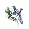
| ||||||||
|---|---|---|---|---|---|---|---|---|---|
| 1 |
| ||||||||
| Unit cell |
|
-Components
| #1: Protein | Mass: 10546.639 Da / Num. of mol.: 1 Source method: isolated from a genetically manipulated source Source: (gene. exp.) Vaccinia virus (strain Western Reserve)Strain: Western Reserve / Gene: VACWR079, G3L / Production host:  |
|---|---|
| #2: Protein | Mass: 9221.222 Da / Num. of mol.: 1 Source method: isolated from a genetically manipulated source Source: (gene. exp.) Vaccinia virus (strain Western Reserve)Strain: Western Reserve / Gene: VACWR092, L5R / Production host: |
| #3: Water | ChemComp-HOH /  Mass: 18.015 Da / Num. of mol.: 62 / Source method: isolated from a natural source / Formula: H2O Mass: 18.015 Da / Num. of mol.: 62 / Source method: isolated from a natural source / Formula: H2O |
| Has ligand of interest | N |
| Has protein modification | Y |
-Experimental details
-Experiment
| Experiment | Method: X-RAY DIFFRACTION / Number of used crystals: 1 |
|---|
- Sample preparation
| Crystal grow | Temperature: 291 K / Method: vapor diffusion, sitting drop / pH: 6.2 Details: 0.2 M Ammonium nitrate (pH 6.2), 20% w/v Polyethylene glycol 3350 |
|---|
-Data collection
| Diffraction | Mean temperature: 100 K / Serial crystal experiment: N |
|---|---|
| Diffraction source | Source: SYNCHROTRON / Site: SSRF  / Beamline: BL19U1 / Wavelength: 0.97892 Å / Beamline: BL19U1 / Wavelength: 0.97892 Å |
| Detector | Type: DECTRIS PILATUS3 S 6M / Detector: PIXEL / Date: Oct 5, 2018 |
| Radiation | Protocol: SINGLE WAVELENGTH / Monochromatic (M) / Laue (L): M / Scattering type: x-ray |
| Radiation wavelength | Wavelength: 0.97892 Å / Relative weight: 1 |
| Reflection | Resolution: 1.8→50 Å / Num. obs: 11689 / % possible obs: 99.4 % / Redundancy: 5.2 % / Rmerge(I) obs: 0.113 / Net I/σ(I): 15.87 |
| Reflection shell | Resolution: 1.8→1.86 Å / Redundancy: 4.8 % / Rmerge(I) obs: 0.544 / Mean I/σ(I) obs: 2 / Num. unique obs: 1143 / % possible all: 98.8 |
- Processing
| Software |
| ||||||||||||||||||||||||||||||||||||||||
|---|---|---|---|---|---|---|---|---|---|---|---|---|---|---|---|---|---|---|---|---|---|---|---|---|---|---|---|---|---|---|---|---|---|---|---|---|---|---|---|---|---|
| Refinement | Method to determine structure: SAD / Resolution: 1.81→29.248 Å / SU ML: 0.17 / Cross valid method: THROUGHOUT / σ(F): 1.4 / Phase error: 27.26 / Stereochemistry target values: ML
| ||||||||||||||||||||||||||||||||||||||||
| Solvent computation | Shrinkage radii: 0.9 Å / VDW probe radii: 1.11 Å / Solvent model: FLAT BULK SOLVENT MODEL | ||||||||||||||||||||||||||||||||||||||||
| Displacement parameters | Biso max: 102.48 Å2 / Biso mean: 39.9118 Å2 / Biso min: 19.54 Å2 | ||||||||||||||||||||||||||||||||||||||||
| Refinement step | Cycle: final / Resolution: 1.81→29.248 Å
| ||||||||||||||||||||||||||||||||||||||||
| Refine LS restraints |
| ||||||||||||||||||||||||||||||||||||||||
| LS refinement shell | Refine-ID: X-RAY DIFFRACTION / Rfactor Rfree error: 0
| ||||||||||||||||||||||||||||||||||||||||
| Refinement TLS params. | Method: refined / Origin x: 4.3902 Å / Origin y: 1.6321 Å / Origin z: 16.1136 Å
| ||||||||||||||||||||||||||||||||||||||||
| Refinement TLS group | Selection details: all |
