 Movie
Movie Controller


+ Open data

- Basic information
| Entry | Database: PDB / ID: 7ytf | ||||||
|---|---|---|---|---|---|---|---|
| Title | Structure of OCPx2 from Nostoc flagelliforme CCNUN1 | ||||||
 Components Components | Ketosteroid isomerase-related protein | ||||||
 Keywords Keywords | PHOTOSYNTHESIS / OCP protein | ||||||
| Function / homology |  Function and homology information Function and homology informationlight absorption / phycobilisome / chloride ion binding / isomerase activity Similarity search - Function | ||||||
| Biological species |  Nostoc flagelliforme CCNUN1 (bacteria) Nostoc flagelliforme CCNUN1 (bacteria) | ||||||
| Method |  X-RAY DIFFRACTION / SYNCHROTRON / MOLECULAR REPLACEMENT / Resolution: 2.65 Å X-RAY DIFFRACTION / SYNCHROTRON / MOLECULAR REPLACEMENT / Resolution: 2.65 Å | ||||||
 Authors Authors | Yang, Y.W. / Chen, S.Z. / Liu, K. / Chen, M. / Qiu, B.S. | ||||||
| Funding support |  China, 1items China, 1items
| ||||||
 Citation Citation | Journal: Plant Physiol. / Year: 2023 Title: Functional specialization of expanded orange carotenoid protein paralogs in subaerial Nostoc species. Authors: Yang, Y.W. / Liu, K. / Huang, D. / Yu, C. / Chen, S.Z. / Chen, M. / Qiu, B.S. | ||||||
| History |
|
- Structure visualization
| Structure viewer | Molecule: MolmilJmol/JSmol |
|---|
- Downloads & links
-Download
| PDBx/mmCIF format | 7ytf.cif.gz | 78.2 KB | Display | PDBx/mmCIF format |
|---|---|---|---|---|
| PDB format | pdb7ytf.ent.gz | 56.4 KB | Display | PDB format |
| PDBx/mmJSON format | 7ytf.json.gz | Tree view | PDBx/mmJSON format | |
| Others |  Other downloads Other downloads |
-Validation report
| Arichive directory | https://data.pdbj.org/pub/pdb/validation_reports/yt/7ytfftp://data.pdbj.org/pub/pdb/validation_reports/yt/7ytf | HTTPS FTP |
|---|
-Related structure data
| Related structure data | 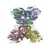 7ythC  4xb5S S: Starting model for refinement C: citing same article ( |
|---|---|
| Similar structure data |
-Links
 PDBj
PDBj
- Assembly
| Deposited unit | 
| ||||||||
|---|---|---|---|---|---|---|---|---|---|
| 1 |
| ||||||||
| Unit cell |
|
-Components
| #1: Protein | Mass: 36202.066 Da / Num. of mol.: 1 Source method: isolated from a genetically manipulated source Source: (gene. exp.) Nostoc flagelliforme CCNUN1 (bacteria) / Gene: COO91_05528 / Production host: |
|---|---|
| #2: Chemical | ChemComp-45D /   Mass: 564.840 Da / Num. of mol.: 1 / Source method: obtained synthetically / Formula: C40H52O2 / Feature type: SUBJECT OF INVESTIGATION Mass: 564.840 Da / Num. of mol.: 1 / Source method: obtained synthetically / Formula: C40H52O2 / Feature type: SUBJECT OF INVESTIGATION |
| #3: Water | ChemComp-HOH /  Mass: 18.015 Da / Num. of mol.: 52 / Source method: isolated from a natural source / Formula: H2O Mass: 18.015 Da / Num. of mol.: 52 / Source method: isolated from a natural source / Formula: H2O |
| Has ligand of interest | Y |
-Experimental details
-Experiment
| Experiment | Method: X-RAY DIFFRACTION / Number of used crystals: 1 |
|---|
- Sample preparation
| Crystal | Density Matthews: 4.31 Å3/Da / Density % sol: 71.49 % |
|---|---|
| Crystal grow | Temperature: 291 K / Method: vapor diffusion, sitting drop / Details: 0.1 M BIS-TRIS pH 5.5, 2.0 M Ammonium sulfate |
-Data collection
| Diffraction | Mean temperature: 100 K / Serial crystal experiment: N |
|---|---|
| Diffraction source | Source: SYNCHROTRON / Site: SSRF / Beamline: BL17U1 / Wavelength: 0.979183 Å |
| Detector | Type: DECTRIS EIGER X 16M / Detector: PIXEL / Date: Apr 13, 2018 |
| Radiation | Protocol: SINGLE WAVELENGTH / Monochromatic (M) / Laue (L): M / Scattering type: x-ray |
| Radiation wavelength | Wavelength: 0.979183 Å / Relative weight: 1 |
| Reflection | Resolution: 2.65→66.96 Å / Num. obs: 18058 / % possible obs: 98.6 % / Redundancy: 39.3 % / CC1/2: 0.999 / Rmerge(I) obs: 0.099 / Net I/σ(I): 29.5 |
| Reflection shell | Resolution: 2.65→2.78 Å / Rmerge(I) obs: 0.781 / Mean I/σ(I) obs: 5.9 / Num. unique obs: 2372 / CC1/2: 0.985 / % possible all: 100 |
- Processing
| Software |
| ||||||||||||||||||||||||||||||||||||||||||||||||||||||||||||
|---|---|---|---|---|---|---|---|---|---|---|---|---|---|---|---|---|---|---|---|---|---|---|---|---|---|---|---|---|---|---|---|---|---|---|---|---|---|---|---|---|---|---|---|---|---|---|---|---|---|---|---|---|---|---|---|---|---|---|---|---|---|
| Refinement | Method to determine structure: MOLECULAR REPLACEMENT Starting model: 4XB5 Resolution: 2.65→44.68 Å / Cor.coef. Fo:Fc: 0.953 / Cor.coef. Fo:Fc free: 0.923 / SU B: 8.617 / SU ML: 0.183 / Cross valid method: THROUGHOUT / σ(F): 0 / ESU R: 0.32 / ESU R Free: 0.266 / Stereochemistry target values: MAXIMUM LIKELIHOOD Details: HYDROGENS HAVE BEEN ADDED IN THE RIDING POSITIONS U VALUES : REFINED INDIVIDUALLY
| ||||||||||||||||||||||||||||||||||||||||||||||||||||||||||||
| Solvent computation | Ion probe radii: 0.8 Å / Shrinkage radii: 0.8 Å / VDW probe radii: 1.2 Å / Solvent model: MASK | ||||||||||||||||||||||||||||||||||||||||||||||||||||||||||||
| Displacement parameters | Biso max: 187.16 Å2 / Biso mean: 66.57 Å2 / Biso min: 39.5 Å2
| ||||||||||||||||||||||||||||||||||||||||||||||||||||||||||||
| Refinement step | Cycle: final / Resolution: 2.65→44.68 Å
| ||||||||||||||||||||||||||||||||||||||||||||||||||||||||||||
| Refine LS restraints |
| ||||||||||||||||||||||||||||||||||||||||||||||||||||||||||||
| LS refinement shell | Resolution: 2.65→2.719 Å / Rfactor Rfree error: 0 / Total num. of bins used: 20
|
