 Movie
Movie Controller

[English] 日本語
 Yorodumi
- PDB-7ysl: Crystal structure of D-Cysteine desulfhydrase with a trapped PLP-... -
+ Open data

- Basic information
| Entry | Database: PDB / ID: 7ysl | ||||||||||||
|---|---|---|---|---|---|---|---|---|---|---|---|---|---|
| Title | Crystal structure of D-Cysteine desulfhydrase with a trapped PLP-pyruvate geminal diamine | ||||||||||||
 Components Components | D-Cysteine desulfhydrase | ||||||||||||
 Keywords Keywords | LYASE / PLP-dependent enzyme / D-Cysteine desulfhydrase / geminal diamine | ||||||||||||
| Function / homology |  Function and homology information Function and homology informationL-cysteate sulfo-lyase / 1-aminocyclopropane-1-carboxylate deaminase activity / D-cysteine desulfhydrase activity Similarity search - Function | ||||||||||||
| Biological species |  Pectobacterium atrosepticum SCRI1043 (bacteria) Pectobacterium atrosepticum SCRI1043 (bacteria) | ||||||||||||
| Method |  X-RAY DIFFRACTION / SYNCHROTRON / MOLECULAR REPLACEMENT / Resolution: 2.02 Å X-RAY DIFFRACTION / SYNCHROTRON / MOLECULAR REPLACEMENT / Resolution: 2.02 Å | ||||||||||||
 Authors Authors | Zhang, X. / Wang, L. / Xu, X. / Xing, X. / Zhou, J. | ||||||||||||
| Funding support |  China, 1items China, 1items
| ||||||||||||
 Citation Citation | Journal: Tetrahedron / Year: 2022 Title: Characterization and structural basis of D-cysteine desulfhydrase from Pectobacterium atrosepticum Authors: Xu, X. / Yang, L. / Zhang, X. / Xing, X. / Zhou, J. | ||||||||||||
| History |
|
- Structure visualization
| Structure viewer | Molecule: MolmilJmol/JSmol |
|---|
- Downloads & links
-Download
| PDBx/mmCIF format | 7ysl.cif.gz | 152.4 KB | Display | PDBx/mmCIF format |
|---|---|---|---|---|
| PDB format | pdb7ysl.ent.gz | 116.2 KB | Display | PDB format |
| PDBx/mmJSON format | 7ysl.json.gz | Tree view | PDBx/mmJSON format | |
| Others |  Other downloads Other downloads |
-Validation report
| Summary document | 7ysl_validation.pdf.gz | 465.7 KB | Display | wwPDB validaton report |
|---|---|---|---|---|
| Full document | 7ysl_full_validation.pdf.gz | 469.6 KB | Display | |
| Data in XML | 7ysl_validation.xml.gz | 28.9 KB | Display | |
| Data in CIF | 7ysl_validation.cif.gz | 40.9 KB | Display | |
| Arichive directory | https://data.pdbj.org/pub/pdb/validation_reports/ys/7yslftp://data.pdbj.org/pub/pdb/validation_reports/ys/7ysl | HTTPS FTP |
-Related structure data
| Related structure data | 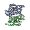 7yskC  4d97S C: citing same article ( S: Starting model for refinement |
|---|---|
| Similar structure data |
-Links
 PDBj
PDBj
- Assembly
| Deposited unit | 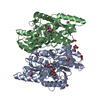
| ||||||||
|---|---|---|---|---|---|---|---|---|---|
| 1 |
| ||||||||
| Unit cell |
|
-Components
| #1: Protein | Mass: 40052.246 Da / Num. of mol.: 2 Source method: isolated from a genetically manipulated source Source: (gene. exp.) Pectobacterium atrosepticum SCRI1043 (bacteria)Strain: SCRI 1043 / ATCC BAA-672 / Gene: ECA1531 / Production host: #2: Chemical | ChemComp-FMT /   Mass: 46.025 Da / Num. of mol.: 12 / Source method: obtained synthetically / Formula: CH2O2 Mass: 46.025 Da / Num. of mol.: 12 / Source method: obtained synthetically / Formula: CH2O2#3: Chemical |   Mass: 62.068 Da / Num. of mol.: 3 / Source method: obtained synthetically / Formula: C2H6O2 Mass: 62.068 Da / Num. of mol.: 3 / Source method: obtained synthetically / Formula: C2H6O2#4: Water | ChemComp-HOH / |  Mass: 18.015 Da / Num. of mol.: 311 / Source method: isolated from a natural source / Formula: H2O Mass: 18.015 Da / Num. of mol.: 311 / Source method: isolated from a natural source / Formula: H2OHas ligand of interest | Y | |
|---|
-Experimental details
-Experiment
| Experiment | Method: X-RAY DIFFRACTION / Number of used crystals: 1 |
|---|
- Sample preparation
| Crystal | Density Matthews: 2.46 Å3/Da / Density % sol: 49.93 % |
|---|---|
| Crystal grow | Temperature: 289.15 K / Method: vapor diffusion, sitting drop Details: 0.1 M Carboxylic acids (sodium formate, ammonium acetate, sodium citrate, potassium sodium tartrate, sodium oxamate), 0.1 M imidazole/MES pH 6.5, 30% (v/v) PEG 500 MME/PEG 20000 |
-Data collection
| Diffraction | Mean temperature: 80 K / Serial crystal experiment: N |
|---|---|
| Diffraction source | Source: SYNCHROTRON / Site: SSRF / Beamline: BL19U1 / Wavelength: 0.9785 Å |
| Detector | Type: DECTRIS PILATUS3 6M / Detector: PIXEL / Date: Jan 16, 2022 |
| Radiation | Protocol: SINGLE WAVELENGTH / Monochromatic (M) / Laue (L): M / Scattering type: x-ray |
| Radiation wavelength | Wavelength: 0.9785 Å / Relative weight: 1 |
| Reflection | Resolution: 2.02→123.09 Å / Num. obs: 46857 / % possible obs: 97.4 % / Redundancy: 7.5 % / Biso Wilson estimate: 28.55 Å2 / CC1/2: 0.998 / Rmerge(I) obs: 0.106 / Net I/σ(I): 13 |
| Reflection shell | Resolution: 2.02→2.07 Å / Redundancy: 7 % / Rmerge(I) obs: 0.92 / Mean I/σ(I) obs: 2.1 / Num. unique obs: 3518 / CC1/2: 0.746 / % possible all: 99.8 |
- Processing
| Software |
| |||||||||||||||||||||||||||||||||||||||||||||||||||||||||||||||||||||||||||||||||||||||||||||||||||||||||||||||||||||||
|---|---|---|---|---|---|---|---|---|---|---|---|---|---|---|---|---|---|---|---|---|---|---|---|---|---|---|---|---|---|---|---|---|---|---|---|---|---|---|---|---|---|---|---|---|---|---|---|---|---|---|---|---|---|---|---|---|---|---|---|---|---|---|---|---|---|---|---|---|---|---|---|---|---|---|---|---|---|---|---|---|---|---|---|---|---|---|---|---|---|---|---|---|---|---|---|---|---|---|---|---|---|---|---|---|---|---|---|---|---|---|---|---|---|---|---|---|---|---|---|---|
| Refinement | Method to determine structure: MOLECULAR REPLACEMENT Starting model: 4D97 Resolution: 2.02→70.21 Å / SU ML: 0.19 / Cross valid method: THROUGHOUT / σ(F): 1.34 / Phase error: 20.76 / Stereochemistry target values: ML
| |||||||||||||||||||||||||||||||||||||||||||||||||||||||||||||||||||||||||||||||||||||||||||||||||||||||||||||||||||||||
| Solvent computation | Shrinkage radii: 0.9 Å / VDW probe radii: 1.1 Å / Solvent model: FLAT BULK SOLVENT MODEL | |||||||||||||||||||||||||||||||||||||||||||||||||||||||||||||||||||||||||||||||||||||||||||||||||||||||||||||||||||||||
| Displacement parameters | Biso max: 101 Å2 / Biso mean: 31.1538 Å2 / Biso min: 15.19 Å2 | |||||||||||||||||||||||||||||||||||||||||||||||||||||||||||||||||||||||||||||||||||||||||||||||||||||||||||||||||||||||
| Refinement step | Cycle: final / Resolution: 2.02→70.21 Å
| |||||||||||||||||||||||||||||||||||||||||||||||||||||||||||||||||||||||||||||||||||||||||||||||||||||||||||||||||||||||
| LS refinement shell | Refine-ID: X-RAY DIFFRACTION / Rfactor Rfree error: 0 / Total num. of bins used: 16
|
