 Movie
Movie Controller


+ Open data

- Basic information
| Entry | Database: PDB / ID: 7yro | ||||||
|---|---|---|---|---|---|---|---|
| Title | Crystal structure of mango fucosyltransferase 13 | ||||||
 Components Components | Fucosyltransferase | ||||||
 Keywords Keywords | TRANSFERASE / fucosyltransferase / FUT13 / mango | ||||||
| Function / homology |  Function and homology information Function and homology informationalpha-(1->3)-fucosyltransferase activity / : / Transferases; Glycosyltransferases; Hexosyltransferases / Golgi cisterna membrane / : Similarity search - Function | ||||||
| Biological species |  Mangifera indica (mango) Mangifera indica (mango) | ||||||
| Method |  X-RAY DIFFRACTION / SYNCHROTRON / MOLECULAR REPLACEMENT / Resolution: 2.42 Å X-RAY DIFFRACTION / SYNCHROTRON / MOLECULAR REPLACEMENT / Resolution: 2.42 Å | ||||||
 Authors Authors | Okada, T. / Teramoto, T. / Ihara, H. / Ikeda, Y. / Kakuta, Y. | ||||||
| Funding support |  Japan, 1items Japan, 1items
| ||||||
 Citation Citation | Journal: Glycobiology / Year: 2024 Title: Crystal structure of mango alpha 1,3/ alpha 1,4-fucosyltransferase elucidates unique elements that regulate Lewis A-dominant oligosaccharide assembly. Authors: Okada, T. / Teramoto, T. / Ihara, H. / Ikeda, Y. / Kakuta, Y. | ||||||
| History |
|
- Structure visualization
| Structure viewer | Molecule: MolmilJmol/JSmol |
|---|
- Downloads & links
-Download
| PDBx/mmCIF format | 7yro.cif.gz | 541.2 KB | Display | PDBx/mmCIF format |
|---|---|---|---|---|
| PDB format | pdb7yro.ent.gz | 442.7 KB | Display | PDB format |
| PDBx/mmJSON format | 7yro.json.gz | Tree view | PDBx/mmJSON format | |
| Others |  Other downloads Other downloads |
-Validation report
| Summary document | 7yro_validation.pdf.gz | 10.7 MB | Display | wwPDB validaton report |
|---|---|---|---|---|
| Full document | 7yro_full_validation.pdf.gz | 10.7 MB | Display | |
| Data in XML | 7yro_validation.xml.gz | 97.4 KB | Display | |
| Data in CIF | 7yro_validation.cif.gz | 131.1 KB | Display | |
| Arichive directory | https://data.pdbj.org/pub/pdb/validation_reports/yr/7yroftp://data.pdbj.org/pub/pdb/validation_reports/yr/7yro | HTTPS FTP |
-Related structure data
| Similar structure data |
|---|
-Links
 PDBj
PDBj

- Assembly
| Deposited unit | 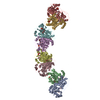
| ||||||||
|---|---|---|---|---|---|---|---|---|---|
| 1 | 
| ||||||||
| 2 | 
| ||||||||
| 3 | 
| ||||||||
| 4 | 
| ||||||||
| Unit cell |
|
-Components
-Protein / Sugars , 2 types, 13 molecules ABCDEFGH
| #1: Protein | Mass: 46161.164 Da / Num. of mol.: 8 Source method: isolated from a genetically manipulated source Source: (gene. exp.) Mangifera indica (mango) / Gene: FUT13 / Production host:   Spodoptera frugiperda (fall armyworm) Spodoptera frugiperda (fall armyworm)References: UniProt: A0A292GKJ7, Transferases; Glycosyltransferases; Hexosyltransferases #6: Sugar | ChemComp-NAG /  Type: D-saccharide, beta linking / Mass: 221.208 Da / Num. of mol.: 5 Type: D-saccharide, beta linking / Mass: 221.208 Da / Num. of mol.: 5Source method: isolated from a genetically manipulated source Formula: C8H15NO6 / Feature type: SUBJECT OF INVESTIGATION |
|---|
-Non-polymers , 5 types, 507 molecules 


| #2: Chemical | ChemComp-GOL /  Mass: 92.094 Da / Num. of mol.: 21 / Source method: obtained synthetically / Formula: C3H8O3 / Feature type: SUBJECT OF INVESTIGATION Mass: 92.094 Da / Num. of mol.: 21 / Source method: obtained synthetically / Formula: C3H8O3 / Feature type: SUBJECT OF INVESTIGATION#3: Chemical | ChemComp-ACY /  Mass: 60.052 Da / Num. of mol.: 7 / Source method: obtained synthetically / Formula: C2H4O2 / Feature type: SUBJECT OF INVESTIGATION Mass: 60.052 Da / Num. of mol.: 7 / Source method: obtained synthetically / Formula: C2H4O2 / Feature type: SUBJECT OF INVESTIGATION#4: Chemical | ChemComp-PO4 / |  Mass: 94.971 Da / Num. of mol.: 1 / Source method: obtained synthetically / Formula: PO4 / Feature type: SUBJECT OF INVESTIGATION Mass: 94.971 Da / Num. of mol.: 1 / Source method: obtained synthetically / Formula: PO4 / Feature type: SUBJECT OF INVESTIGATION#5: Chemical | ChemComp-PPV /  Mass: 177.975 Da / Num. of mol.: 7 / Source method: obtained synthetically / Formula: H4O7P2 / Feature type: SUBJECT OF INVESTIGATION Mass: 177.975 Da / Num. of mol.: 7 / Source method: obtained synthetically / Formula: H4O7P2 / Feature type: SUBJECT OF INVESTIGATION#7: Water | ChemComp-HOH / | Mass: 18.015 Da / Num. of mol.: 471 / Source method: isolated from a natural source / Formula: H2O |
|---|
-Details
| Has ligand of interest | Y |
|---|---|
| Has protein modification | Y |
-Experimental details
-Experiment
| Experiment | Method: X-RAY DIFFRACTION / Number of used crystals: 1 |
|---|
- Sample preparation
| Crystal | Density Matthews: 2.64 Å3/Da / Density % sol: 53.41 % |
|---|---|
| Crystal grow | Temperature: 293.15 K / Method: vapor diffusion, sitting drop Details: 0.2 M Calcium acetate hydrate and 20% w/v Polyethylene glycol 3,350 |
-Data collection
| Diffraction | Mean temperature: 100 K / Serial crystal experiment: N |
|---|---|
| Diffraction source | Source: SYNCHROTRON / Site: SPring-8 / Beamline: BL45XU / Wavelength: 1 Å |
| Detector | Type: DECTRIS PILATUS3 6M / Detector: PIXEL / Date: May 15, 2021 |
| Radiation | Protocol: SINGLE WAVELENGTH / Monochromatic (M) / Laue (L): M / Scattering type: x-ray |
| Radiation wavelength | Wavelength: 1 Å / Relative weight: 1 |
| Reflection | Resolution: 2.42→50 Å / Num. obs: 149646 / % possible obs: 100 % / Redundancy: 9.78 % / CC1/2: 0.995 / Net I/σ(I): 8.55 |
| Reflection shell | Resolution: 2.42→2.56 Å / Mean I/σ(I) obs: 1.14 / Num. unique obs: 24409 / CC1/2: 0.544 |
- Processing
| Software |
| |||||||||||||||||||||||||||||||||||||||||||||||||||||||||||||||||||||||||||||||||||||||||||||||||||||||||
|---|---|---|---|---|---|---|---|---|---|---|---|---|---|---|---|---|---|---|---|---|---|---|---|---|---|---|---|---|---|---|---|---|---|---|---|---|---|---|---|---|---|---|---|---|---|---|---|---|---|---|---|---|---|---|---|---|---|---|---|---|---|---|---|---|---|---|---|---|---|---|---|---|---|---|---|---|---|---|---|---|---|---|---|---|---|---|---|---|---|---|---|---|---|---|---|---|---|---|---|---|---|---|---|---|---|---|
| Refinement | Method to determine structure: MOLECULAR REPLACEMENT Starting model: predicted model Resolution: 2.42→46.86 Å / SU ML: 0.35 / Cross valid method: FREE R-VALUE / σ(F): 1.35 / Phase error: 27.51 / Stereochemistry target values: ML
| |||||||||||||||||||||||||||||||||||||||||||||||||||||||||||||||||||||||||||||||||||||||||||||||||||||||||
| Solvent computation | Shrinkage radii: 0.9 Å / VDW probe radii: 1.11 Å / Solvent model: FLAT BULK SOLVENT MODEL | |||||||||||||||||||||||||||||||||||||||||||||||||||||||||||||||||||||||||||||||||||||||||||||||||||||||||
| Refinement step | Cycle: LAST / Resolution: 2.42→46.86 Å
| |||||||||||||||||||||||||||||||||||||||||||||||||||||||||||||||||||||||||||||||||||||||||||||||||||||||||
| Refine LS restraints |
| |||||||||||||||||||||||||||||||||||||||||||||||||||||||||||||||||||||||||||||||||||||||||||||||||||||||||
| LS refinement shell |
|
