 Movie
Movie Controller


+ Open data

- Basic information
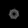
| Entry | Database: PDB / ID: 7yqc | |||||||||||||||||||||||||||||||||
|---|---|---|---|---|---|---|---|---|---|---|---|---|---|---|---|---|---|---|---|---|---|---|---|---|---|---|---|---|---|---|---|---|---|---|
| Title | EM structure of human PA28gamma | |||||||||||||||||||||||||||||||||
 Components Components | Proteasome activator complex subunit 3 | |||||||||||||||||||||||||||||||||
 Keywords Keywords | PROTEIN TRANSPORT / proteasome activator gamma / PA28gamma / PSME3 | |||||||||||||||||||||||||||||||||
| Function / homology |  Function and homology information Function and homology informationproteasome activator complex / Proteasome assembly / regulation of G1/S transition of mitotic cell cycle / endopeptidase activator activity / regulation of proteasomal protein catabolic process / MDM2/MDM4 family protein binding / proteasome complex / negative regulation of extrinsic apoptotic signaling pathway / p53 binding / cilium ...proteasome activator complex / Proteasome assembly / regulation of G1/S transition of mitotic cell cycle / endopeptidase activator activity / regulation of proteasomal protein catabolic process / MDM2/MDM4 family protein binding / proteasome complex / negative regulation of extrinsic apoptotic signaling pathway / p53 binding / cilium / ciliary basal body / apoptotic process / nucleoplasm / identical protein binding / nucleus / membrane / cytosol / cytoplasm Similarity search - Function | |||||||||||||||||||||||||||||||||
| Biological species |  Homo sapiens (human) Homo sapiens (human) | |||||||||||||||||||||||||||||||||
| Method | ELECTRON MICROSCOPY / single particle reconstruction / cryo EM / Resolution: 2.82 Å | |||||||||||||||||||||||||||||||||
 Authors Authors | Chen, D.-D. / Hao, J. / Shen, C.-H. / Yun, C.-H. | |||||||||||||||||||||||||||||||||
| Funding support |  China, 1items China, 1items
| |||||||||||||||||||||||||||||||||
 Citation Citation | Journal: Int J Biol Macromol / Year: 2022 Title: Atomic resolution Cryo-EM structure of human proteasome activator PA28γ. Authors: Dan-Dan Chen / Jia Hao / Chao-Hui Shen / Xian-Ming Deng / Cai-Hong Yun / Abstract: The PA28 family proteasome activators play important roles in regulating proteasome activities. Though the three paralogs (PA28α, PA28β, and PA28γ) are similar in terms of primary sequence, they ...The PA28 family proteasome activators play important roles in regulating proteasome activities. Though the three paralogs (PA28α, PA28β, and PA28γ) are similar in terms of primary sequence, they show significant differences in expression pattern, cellular localization and most importantly, biological functions. While PA28αβ is responsible for promoting peptidase activity of proteasome to facilitate MHC-I antigen processing, but unable to promote protein degradation, PA28γ is well-known to not only promote peptidase activity but also proteolytic activity of proteasome. However, why this paralog has the unique function remains elusive. Previous structural studies have mainly focused on mammalian PA28α, PA28β and PA28αβ heptamers, while structural studies on mammalian PA28γ of atomic resolution are still absent to date. In the present work, we determined the Cryo-EM structure of the human PA28γ heptamer at atomic resolution, revealing interesting unique structural features that may hint our understanding the functional mechanisms of this proteasome activator. | |||||||||||||||||||||||||||||||||
| History |
|
- Structure visualization
| Structure viewer | Molecule: MolmilJmol/JSmol |
|---|
- Downloads & links
-Download
| PDBx/mmCIF format | 7yqc.cif.gz | 251.4 KB | Display | PDBx/mmCIF format |
|---|---|---|---|---|
| PDB format | pdb7yqc.ent.gz | 204.5 KB | Display | PDB format |
| PDBx/mmJSON format | 7yqc.json.gz | Tree view | PDBx/mmJSON format | |
| Others |  Other downloads Other downloads |
-Validation report
| Summary document | 7yqc_validation.pdf.gz | 1.2 MB | Display | wwPDB validaton report |
|---|---|---|---|---|
| Full document | 7yqc_full_validation.pdf.gz | 1.2 MB | Display | |
| Data in XML | 7yqc_validation.xml.gz | 44.4 KB | Display | |
| Data in CIF | 7yqc_validation.cif.gz | 66.8 KB | Display | |
| Arichive directory | https://data.pdbj.org/pub/pdb/validation_reports/yq/7yqcftp://data.pdbj.org/pub/pdb/validation_reports/yq/7yqc | HTTPS FTP |
-Related structure data
| Related structure data | 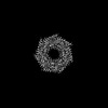 34023MC 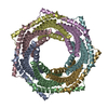 7yqdC M: map data used to model this data C: citing same article ( |
|---|---|
| Similar structure data |
-Links
 PDBj
PDBj

- Assembly
| Deposited unit | 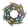
|
|---|---|
| 1 |
|
-Components
| #1: Protein | Mass: 29432.904 Da / Num. of mol.: 7 Source method: isolated from a genetically manipulated source Source: (gene. exp.) Homo sapiens (human) / Gene: PSME3 / Production host:  Has protein modification | N | |
|---|
-Experimental details
-Experiment
| Experiment | Method: ELECTRON MICROSCOPY |
|---|---|
| EM experiment | Aggregation state: PARTICLE / 3D reconstruction method: single particle reconstruction |
- Sample preparation
| Component | Name: Proteasome Activator / Type: ORGANELLE OR CELLULAR COMPONENT / Entity ID: all / Source: RECOMBINANT |
|---|---|
| Source (natural) | Organism: Homo sapiens (human) |
| Source (recombinant) | Organism: |
| Buffer solution | pH: 8 |
| Specimen | Embedding applied: NO / Shadowing applied: NO / Staining applied: NO / Vitrification applied: YES |
| Vitrification | Cryogen name: ETHANE |
- Electron microscopy imaging
| Experimental equipment |  Model: Titan Krios / Image courtesy: FEI Company |
|---|---|
| Microscopy | Model: FEI TITAN KRIOS |
| Electron gun | Electron source:  FIELD EMISSION GUN / Accelerating voltage: 300 kV / Illumination mode: FLOOD BEAM FIELD EMISSION GUN / Accelerating voltage: 300 kV / Illumination mode: FLOOD BEAM |
| Electron lens | Mode: BRIGHT FIELD / Nominal defocus max: 2000 nm / Nominal defocus min: 1000 nm |
| Image recording | Electron dose: 50 e/Å2 / Film or detector model: GATAN K3 (6k x 4k) |
- Processing
| Software | Name: PHENIX / Version: 1.19.2_4158: / Classification: refinement | ||||||||||||||||||||||||
|---|---|---|---|---|---|---|---|---|---|---|---|---|---|---|---|---|---|---|---|---|---|---|---|---|---|
| EM software | Name: PHENIX / Category: model refinement | ||||||||||||||||||||||||
| CTF correction | Type: NONE | ||||||||||||||||||||||||
| 3D reconstruction | Resolution: 2.82 Å / Resolution method: FSC 0.143 CUT-OFF / Num. of particles: 395286 / Symmetry type: POINT | ||||||||||||||||||||||||
| Refine LS restraints |
|
