 Movie
Movie Controller

[English] 日本語
 Yorodumi
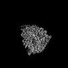
- PDB-7yov: Cryo-EM structure of RNA polymerase in complex with P protein tet... -
+ Open data

- Basic information
| Entry | Database: PDB / ID: 7yov | |||||||||||||||||||||||||||||||||||||||||||||
|---|---|---|---|---|---|---|---|---|---|---|---|---|---|---|---|---|---|---|---|---|---|---|---|---|---|---|---|---|---|---|---|---|---|---|---|---|---|---|---|---|---|---|---|---|---|---|
| Title | Cryo-EM structure of RNA polymerase in complex with P protein tetramer of Newcastle disease virus | |||||||||||||||||||||||||||||||||||||||||||||
 Components Components |
| |||||||||||||||||||||||||||||||||||||||||||||
 Keywords Keywords | VIRUS / cryo-EM / L-P complex / Newcastle disease virus | |||||||||||||||||||||||||||||||||||||||||||||
| Function / homology |  Function and homology information Function and homology informationGDP polyribonucleotidyltransferase / Hydrolases; Acting on acid anhydrides; In phosphorus-containing anhydrides / Transferases; Transferring one-carbon groups; Methyltransferases / virion component / host cell cytoplasm / mRNA 5'-cap (guanine-N7-)-methyltransferase activity / RNA-directed RNA polymerase / RNA-directed RNA polymerase activity / GTPase activity / ATP binding Similarity search - Function | |||||||||||||||||||||||||||||||||||||||||||||
| Biological species |  Avian orthoavulavirus 1 Avian orthoavulavirus 1 | |||||||||||||||||||||||||||||||||||||||||||||
| Method | ELECTRON MICROSCOPY / single particle reconstruction / cryo EM / Resolution: 3.25 Å | |||||||||||||||||||||||||||||||||||||||||||||
 Authors Authors | Chen, Y. / Jingyuan, C. / Xiaoying, F. | |||||||||||||||||||||||||||||||||||||||||||||
| Funding support |  China, 1items China, 1items
| |||||||||||||||||||||||||||||||||||||||||||||
 Citation Citation | Journal: Nat Commun / Year: 2023 Title: Structure of the Newcastle Disease Virus L protein in complex with tetrameric phosphoprotein. Authors: Jingyuan Cong / Xiaoying Feng / Huiling Kang / Wangjun Fu / Lei Wang / Chenlong Wang / Xuemei Li / Yutao Chen / Zihe Rao / Abstract: Newcastle disease virus (NDV) belongs to Paramyxoviridae, which contains lethal human and animal pathogens. NDV RNA genome is replicated and transcribed by a multifunctional 250 kDa RNA-dependent ...Newcastle disease virus (NDV) belongs to Paramyxoviridae, which contains lethal human and animal pathogens. NDV RNA genome is replicated and transcribed by a multifunctional 250 kDa RNA-dependent RNA polymerase (L protein). To date, high-resolution structure of NDV L protein complexed with P protein remains to be elucidated, limiting our understanding of the molecular mechanisms of Paramyxoviridae replication/transcription. Here, we used cryo-EM and enzymatic assays to investigate the structure-function relationship of L-P complex. We found that C-terminal of CD-MTase-CTD module of the atomic-resolution L-P complex conformationally rearranges, and the priming/intrusion loops are likely in RNA elongation conformations different from previous structures. The P protein adopts a unique tetrameric organization and interacts with L protein. Our findings indicate that NDV L-P complex represents elongation state distinct from previous structures. Our work greatly advances the understanding of Paramyxoviridae RNA synthesis, revealing how initiation/elongation alternates, providing clues for identifying therapeutic targets against Paramyxoviridae. | |||||||||||||||||||||||||||||||||||||||||||||
| History |
|
- Structure visualization
| Structure viewer | Molecule: MolmilJmol/JSmol |
|---|
- Downloads & links
-Download
| PDBx/mmCIF format | 7yov.cif.gz | 335.5 KB | Display | PDBx/mmCIF format |
|---|---|---|---|---|
| PDB format | pdb7yov.ent.gz | 249.3 KB | Display | PDB format |
| PDBx/mmJSON format | 7yov.json.gz | Tree view | PDBx/mmJSON format | |
| Others |  Other downloads Other downloads |
-Validation report
| Arichive directory | https://data.pdbj.org/pub/pdb/validation_reports/yo/7yovftp://data.pdbj.org/pub/pdb/validation_reports/yo/7yov | HTTPS FTP |
|---|
-Related structure data
| Related structure data | 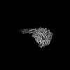 33988MC  7yotC  7youC M: map data used to model this data C: citing same article ( |
|---|---|
| Similar structure data |
-Links
 PDBj
PDBj
- Assembly
| Deposited unit | 
|
|---|---|
| 1 |
|
-Components
| #1: Protein | Mass: 41738.379 Da / Num. of mol.: 4 Source method: isolated from a genetically manipulated source Source: (gene. exp.) Avian orthoavulavirus 1 / Production host:   Spodoptera frugiperda (fall armyworm) / References: UniProt: A0A0S2UXI9 Spodoptera frugiperda (fall armyworm) / References: UniProt: A0A0S2UXI9#2: Protein | | Mass: 249183.188 Da / Num. of mol.: 1 Source method: isolated from a genetically manipulated source Source: (gene. exp.) Avian orthoavulavirus 1 / Production host: Spodoptera frugiperda (fall armyworm)References: UniProt: A0A0S2UX53, RNA-directed RNA polymerase, Hydrolases; Acting on acid anhydrides; In phosphorus-containing anhydrides, GDP polyribonucleotidyltransferase, Transferases; ...References: UniProt: A0A0S2UX53, RNA-directed RNA polymerase, Hydrolases; Acting on acid anhydrides; In phosphorus-containing anhydrides, GDP polyribonucleotidyltransferase, Transferases; Transferring one-carbon groups; Methyltransferases Has protein modification | Y | |
|---|
-Experimental details
-Experiment
| Experiment | Method: ELECTRON MICROSCOPY |
|---|---|
| EM experiment | Aggregation state: 3D ARRAY / 3D reconstruction method: single particle reconstruction |
- Sample preparation
| Component | Name: NDV L-P complex / Type: COMPLEX / Entity ID: all / Source: RECOMBINANT |
|---|---|
| Source (natural) | Organism: Avian orthoavulavirus 1 |
| Source (recombinant) | Organism: Spodoptera frugiperda (fall armyworm) |
| Buffer solution | pH: 8 |
| Specimen | Embedding applied: NO / Shadowing applied: NO / Staining applied: NO / Vitrification applied: YES |
| Vitrification | Cryogen name: ETHANE |
- Electron microscopy imaging
| Experimental equipment |  Model: Titan Krios / Image courtesy: FEI Company |
|---|---|
| Microscopy | Model: FEI TITAN KRIOS |
| Electron gun | Electron source:  FIELD EMISSION GUN / Accelerating voltage: 300 kV / Illumination mode: FLOOD BEAM FIELD EMISSION GUN / Accelerating voltage: 300 kV / Illumination mode: FLOOD BEAM |
| Electron lens | Mode: BRIGHT FIELD / Nominal defocus max: 2500 nm / Nominal defocus min: 1200 nm |
| Image recording | Electron dose: 60 e/Å2 / Film or detector model: GATAN K2 SUMMIT (4k x 4k) |
- Processing
| Software | Name: PHENIX / Version: 1.12_2829: / Classification: refinement |
|---|---|
| EM software | Name: PHENIX / Category: model refinement |
| CTF correction | Type: PHASE FLIPPING ONLY |
| 3D reconstruction | Resolution: 3.25 Å / Resolution method: FSC 0.143 CUT-OFF / Num. of particles: 153000 / Symmetry type: POINT |
