 Movie
Movie Controller


+ Open data

- Basic information
| Entry | Database: PDB / ID: 7yop | ||||||
|---|---|---|---|---|---|---|---|
| Title | Spiroplasma melliferum FtsZ bound to GMPPNP | ||||||
 Components Components | Cell division protein FtsZ | ||||||
 Keywords Keywords | CELL CYCLE / FtsZ / GTP binding / GTPase / cytoskeletal / tubulin homolog | ||||||
| Function / homology | GUANOSINE-5'-DIPHOSPHATE / PHOSPHOAMINOPHOSPHONIC ACID-GUANYLATE ESTER / :  Function and homology information Function and homology information | ||||||
| Biological species |  Spiroplasma melliferum KC3 (bacteria) Spiroplasma melliferum KC3 (bacteria) | ||||||
| Method |  X-RAY DIFFRACTION / SYNCHROTRON / MOLECULAR REPLACEMENT / Resolution: 2.20003123654 Å X-RAY DIFFRACTION / SYNCHROTRON / MOLECULAR REPLACEMENT / Resolution: 2.20003123654 Å | ||||||
 Authors Authors | Chakraborty, J. / Pananghat, G. | ||||||
| Funding support |  India, 1items India, 1items
| ||||||
 Citation Citation | Journal: J.Biol.Chem. / Year: 2024 Title: Dynamics of interdomain rotation facilitates FtsZ filament assembly. Authors: Chakraborty, J. / Poddar, S. / Dutta, S. / Bahulekar, V. / Harne, S. / Srinivasan, R. / Gayathri, P. | ||||||
| History |
|
- Structure visualization
| Structure viewer | Molecule: MolmilJmol/JSmol |
|---|
- Downloads & links
-Download
| PDBx/mmCIF format | 7yop.cif.gz | 157.5 KB | Display | PDBx/mmCIF format |
|---|---|---|---|---|
| PDB format | pdb7yop.ent.gz | 98.6 KB | Display | PDB format |
| PDBx/mmJSON format | 7yop.json.gz | Tree view | PDBx/mmJSON format | |
| Others |  Other downloads Other downloads |
-Validation report
| Arichive directory | https://data.pdbj.org/pub/pdb/validation_reports/yo/7yopftp://data.pdbj.org/pub/pdb/validation_reports/yo/7yop | HTTPS FTP |
|---|
-Related structure data
| Related structure data |  7yszC  8grwC  2vxyS S: Starting model for refinement C: citing same article ( |
|---|---|
| Similar structure data |
-Links
 PDBj
PDBj


- Assembly
| Deposited unit | 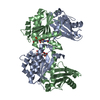
| ||||||||||||
|---|---|---|---|---|---|---|---|---|---|---|---|---|---|
| 1 |
| ||||||||||||
| Unit cell |
| ||||||||||||
| Components on special symmetry positions |
|
-Components
| #1: Protein | Mass: 33630.055 Da / Num. of mol.: 2 Source method: isolated from a genetically manipulated source Source: (gene. exp.) Spiroplasma melliferum KC3 (bacteria) / Gene: ftsZ, SPM_000165 / Production host: #2: Chemical | ChemComp-TRS / |   Mass: 122.143 Da / Num. of mol.: 1 / Source method: obtained synthetically / Formula: C4H12NO3 / Comment: pH buffer*YM Mass: 122.143 Da / Num. of mol.: 1 / Source method: obtained synthetically / Formula: C4H12NO3 / Comment: pH buffer*YM#3: Chemical | ChemComp-GDP / |   Type: RNA linking / Mass: 443.201 Da / Num. of mol.: 1 / Source method: obtained synthetically / Formula: C10H15N5O11P2 / Feature type: SUBJECT OF INVESTIGATION / Comment: GDP, energy-carrying molecule*YM Type: RNA linking / Mass: 443.201 Da / Num. of mol.: 1 / Source method: obtained synthetically / Formula: C10H15N5O11P2 / Feature type: SUBJECT OF INVESTIGATION / Comment: GDP, energy-carrying molecule*YM#4: Chemical | ChemComp-GNP / |   Mass: 522.196 Da / Num. of mol.: 1 / Source method: isolated from a natural source / Formula: C10H17N6O13P3 / Feature type: SUBJECT OF INVESTIGATION Mass: 522.196 Da / Num. of mol.: 1 / Source method: isolated from a natural source / Formula: C10H17N6O13P3 / Feature type: SUBJECT OF INVESTIGATIONComment: GppNHp, GMPPNP, energy-carrying molecule analogue*YM #5: Water | ChemComp-HOH / |  Mass: 18.015 Da / Num. of mol.: 342 / Source method: isolated from a natural source / Formula: H2O Mass: 18.015 Da / Num. of mol.: 342 / Source method: isolated from a natural source / Formula: H2OHas ligand of interest | Y | |
|---|
-Experimental details
-Experiment
| Experiment | Method: X-RAY DIFFRACTION / Number of used crystals: 1 |
|---|
- Sample preparation
| Crystal | Density Matthews: 2.96 Å3/Da / Density % sol: 58.45 % / Description: Needle shape crystals |
|---|---|
| Crystal grow | Temperature: 291.15 K / Method: vapor diffusion, sitting drop Details: 0.3M sodium acetate, 0.1M Tris pH 8.5, 15% w/v PEG 4000 |
-Data collection
| Diffraction | Mean temperature: 100 K / Serial crystal experiment: N |
|---|---|
| Diffraction source | Source: SYNCHROTRON / Site: Diamond  / Beamline: I04 / Wavelength: 0.97623 Å / Beamline: I04 / Wavelength: 0.97623 Å |
| Detector | Type: DECTRIS PILATUS 6M-F / Detector: PIXEL / Date: Dec 10, 2018 |
| Radiation | Protocol: SINGLE WAVELENGTH / Monochromatic (M) / Laue (L): M / Scattering type: x-ray |
| Radiation wavelength | Wavelength: 0.97623 Å / Relative weight: 1 |
| Reflection | Resolution: 2.2→49.2361734647 Å / Num. obs: 38294 / % possible obs: 99.5 % / Redundancy: 5.8 % / Biso Wilson estimate: 33.3678269305 Å2 / CC1/2: 0.998 / Rmerge(I) obs: 0.068 / Rpim(I) all: 0.031 / Rrim(I) all: 0.075 / Net I/σ(I): 17 |
| Reflection shell | Resolution: 2.2→2.2 Å / Redundancy: 5.6 % / Rmerge(I) obs: 0.44 / Mean I/σ(I) obs: 4.3 / Num. unique obs: 17906 / CC1/2: 0.894 / Rpim(I) all: 0.28 / Rrim(I) all: 0.485 / % possible all: 98.9 |
- Processing
| Software |
| |||||||||||||||||||||||||||||||||||||||||||||||||||||||||||||||||||||||||||||||||||||||||||||||||||||||||
|---|---|---|---|---|---|---|---|---|---|---|---|---|---|---|---|---|---|---|---|---|---|---|---|---|---|---|---|---|---|---|---|---|---|---|---|---|---|---|---|---|---|---|---|---|---|---|---|---|---|---|---|---|---|---|---|---|---|---|---|---|---|---|---|---|---|---|---|---|---|---|---|---|---|---|---|---|---|---|---|---|---|---|---|---|---|---|---|---|---|---|---|---|---|---|---|---|---|---|---|---|---|---|---|---|---|---|
| Refinement | Method to determine structure: MOLECULAR REPLACEMENT Starting model: 2VXY Resolution: 2.20003123654→49.2361734647 Å / SU ML: 0.223321967793 / Cross valid method: FREE R-VALUE / σ(F): 1.37111906751 / Phase error: 22.6612563806 Stereochemistry target values: GeoStd + Monomer Library + CDL v1.2
| |||||||||||||||||||||||||||||||||||||||||||||||||||||||||||||||||||||||||||||||||||||||||||||||||||||||||
| Solvent computation | Shrinkage radii: 0.9 Å / VDW probe radii: 1.11 Å / Solvent model: FLAT BULK SOLVENT MODEL | |||||||||||||||||||||||||||||||||||||||||||||||||||||||||||||||||||||||||||||||||||||||||||||||||||||||||
| Displacement parameters | Biso mean: 43.6665595695 Å2 | |||||||||||||||||||||||||||||||||||||||||||||||||||||||||||||||||||||||||||||||||||||||||||||||||||||||||
| Refinement step | Cycle: LAST / Resolution: 2.20003123654→49.2361734647 Å
| |||||||||||||||||||||||||||||||||||||||||||||||||||||||||||||||||||||||||||||||||||||||||||||||||||||||||
| Refine LS restraints |
| |||||||||||||||||||||||||||||||||||||||||||||||||||||||||||||||||||||||||||||||||||||||||||||||||||||||||
| LS refinement shell |
|
