 Movie
Movie Controller


+ Open data

- Basic information
| Entry | Database: PDB / ID: 7ylt | ||||||
|---|---|---|---|---|---|---|---|
| Title | Structure of a bacteria protein | ||||||
 Components Components | Putative ABC transporter, substrate-binding protein | ||||||
 Keywords Keywords | METAL BINDING PROTEIN / a bacteria protein / TRANSPORT PROTEIN | ||||||
| Function / homology | : / Leucine-binding protein domain / Periplasmic binding protein / Periplasmic binding protein-like I / Putative ABC transporter, substrate-binding protein Function and homology information Function and homology information | ||||||
| Biological species |  Variovorax paradoxus (bacteria) Variovorax paradoxus (bacteria) | ||||||
| Method |  X-RAY DIFFRACTION / SYNCHROTRON / MOLECULAR REPLACEMENT / Resolution: 2.3 Å X-RAY DIFFRACTION / SYNCHROTRON / MOLECULAR REPLACEMENT / Resolution: 2.3 Å | ||||||
 Authors Authors | Zhang, H. / Ma, Y.J. / Yu, G.M. / Li, X.Z. | ||||||
| Funding support |  China, 1items China, 1items
| ||||||
 Citation Citation | Journal: To Be Published Title: Structure of a bacteria protein Authors: Zhang, H. / Ma, Y.J. / Yu, G.M. / Li, X.Z. | ||||||
| History |
|
- Structure visualization
| Structure viewer | Molecule: MolmilJmol/JSmol |
|---|
- Downloads & links
-Download
| PDBx/mmCIF format | 7ylt.cif.gz | 940.8 KB | Display | PDBx/mmCIF format |
|---|---|---|---|---|
| PDB format | pdb7ylt.ent.gz | 652.1 KB | Display | PDB format |
| PDBx/mmJSON format | 7ylt.json.gz | Tree view | PDBx/mmJSON format | |
| Others |  Other downloads Other downloads |
-Validation report
| Arichive directory | https://data.pdbj.org/pub/pdb/validation_reports/yl/7yltftp://data.pdbj.org/pub/pdb/validation_reports/yl/7ylt | HTTPS FTP |
|---|
-Related structure data

-Links
 PDBj
PDBj- Assembly
| Deposited unit | 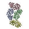
| ||||||||||||
|---|---|---|---|---|---|---|---|---|---|---|---|---|---|
| 1 | 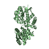
| ||||||||||||
| 2 | 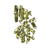
| ||||||||||||
| 3 | 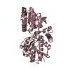
| ||||||||||||
| 4 | 
| ||||||||||||
| Unit cell |
|
-Components
| #1: Protein | Mass: 38375.887 Da / Num. of mol.: 4 Source method: isolated from a genetically manipulated source Source: (gene. exp.) Variovorax paradoxus (bacteria) / Gene: VAPA_1c15920 / Production host: #2: Water | ChemComp-HOH / |  Mass: 18.015 Da / Num. of mol.: 522 / Source method: isolated from a natural source / Formula: H2O Mass: 18.015 Da / Num. of mol.: 522 / Source method: isolated from a natural source / Formula: H2O |
|---|
-Experimental details
-Experiment
| Experiment | Method: X-RAY DIFFRACTION / Number of used crystals: 1 |
|---|
- Sample preparation
| Crystal | Density Matthews: 2.16 Å3/Da / Density % sol: 43.12 % |
|---|---|
| Crystal grow | Temperature: 291.15 K / Method: liquid diffusion Details: Polyethylene glycol 3350, Tris, Cobalt(II) chloride hexahydrate |
-Data collection
| Diffraction | Mean temperature: 100 K / Serial crystal experiment: N |
|---|---|
| Diffraction source | Source: SYNCHROTRON / Site: SSRF / Beamline: BL19U1 / Wavelength: 0.979 Å |
| Detector | Type: DECTRIS PILATUS3 6M / Detector: PIXEL / Date: Oct 6, 2021 |
| Radiation | Protocol: SINGLE WAVELENGTH / Monochromatic (M) / Laue (L): M / Scattering type: x-ray |
| Radiation wavelength | Wavelength: 0.979 Å / Relative weight: 1 |
| Reflection | Resolution: 2.3→48.67 Å / Num. obs: 52101 / % possible obs: 90.99 % / Redundancy: 3.6 % / Biso Wilson estimate: 24.67 Å2 / CC1/2: 0.998 / Net I/σ(I): 9.78 |
| Reflection shell | Resolution: 2.3→2.382 Å / Num. unique obs: 4716 / CC1/2: 0.968 |
- Processing
| Software |
| |||||||||||||||||||||||||||||||||||||||||||||||||||||||||||||||||||||||||||||||||||||||||||||||||||||||||||||||||||||||||||||||||||||
|---|---|---|---|---|---|---|---|---|---|---|---|---|---|---|---|---|---|---|---|---|---|---|---|---|---|---|---|---|---|---|---|---|---|---|---|---|---|---|---|---|---|---|---|---|---|---|---|---|---|---|---|---|---|---|---|---|---|---|---|---|---|---|---|---|---|---|---|---|---|---|---|---|---|---|---|---|---|---|---|---|---|---|---|---|---|---|---|---|---|---|---|---|---|---|---|---|---|---|---|---|---|---|---|---|---|---|---|---|---|---|---|---|---|---|---|---|---|---|---|---|---|---|---|---|---|---|---|---|---|---|---|---|---|---|
| Refinement | Method to determine structure: MOLECULAR REPLACEMENT Starting model: Alphafold2 Resolution: 2.3→48.67 Å / SU ML: 0.2522 / Cross valid method: FREE R-VALUE / σ(F): 1.97 / Phase error: 23.9203 Stereochemistry target values: GeoStd + Monomer Library + CDL v1.2
| |||||||||||||||||||||||||||||||||||||||||||||||||||||||||||||||||||||||||||||||||||||||||||||||||||||||||||||||||||||||||||||||||||||
| Solvent computation | Shrinkage radii: 0.9 Å / VDW probe radii: 1.1 Å / Solvent model: FLAT BULK SOLVENT MODEL | |||||||||||||||||||||||||||||||||||||||||||||||||||||||||||||||||||||||||||||||||||||||||||||||||||||||||||||||||||||||||||||||||||||
| Displacement parameters | Biso mean: 34.07 Å2 | |||||||||||||||||||||||||||||||||||||||||||||||||||||||||||||||||||||||||||||||||||||||||||||||||||||||||||||||||||||||||||||||||||||
| Refinement step | Cycle: LAST / Resolution: 2.3→48.67 Å
| |||||||||||||||||||||||||||||||||||||||||||||||||||||||||||||||||||||||||||||||||||||||||||||||||||||||||||||||||||||||||||||||||||||
| Refine LS restraints |
| |||||||||||||||||||||||||||||||||||||||||||||||||||||||||||||||||||||||||||||||||||||||||||||||||||||||||||||||||||||||||||||||||||||
| LS refinement shell |
| |||||||||||||||||||||||||||||||||||||||||||||||||||||||||||||||||||||||||||||||||||||||||||||||||||||||||||||||||||||||||||||||||||||
| Refinement TLS params. | Method: refined / Origin x: 13.0277649281 Å / Origin y: 19.8693634351 Å / Origin z: 3.99097172731 Å
| |||||||||||||||||||||||||||||||||||||||||||||||||||||||||||||||||||||||||||||||||||||||||||||||||||||||||||||||||||||||||||||||||||||
| Refinement TLS group | Selection details: all |
