 ムービー
ムービー コントローラー


+ データを開く

- 基本情報
基本情報
| 登録情報 | データベース: PDB / ID: 7ylo | ||||||
|---|---|---|---|---|---|---|---|
| タイトル | Conversion of indole-3-acetic acid into indole-3-aldehyde in bacteria Metabolic network of tryptophan around the indole-3-aldehyde formation | ||||||
 要素 要素 | Dyp-type peroxidase | ||||||
 キーワード キーワード | OXIDOREDUCTASE / peroxidase / LyP / BIOSYNTHETIC PROTEIN | ||||||
| 機能・相同性 |  機能・相同性情報 機能・相同性情報酸化還元酵素; 過酸化物を電子受容体にする; ペルオキシダーゼ / peroxidase activity / heme binding / metal ion binding / cytosol 類似検索 - 分子機能 | ||||||
| 生物種 |  Limosilactobacillus fermentum (バクテリア) Limosilactobacillus fermentum (バクテリア) | ||||||
| 手法 |  X線回折 / シンクロトロン / 多重同系置換 / 解像度: 1.83 Å X線回折 / シンクロトロン / 多重同系置換 / 解像度: 1.83 Å | ||||||
 データ登録者 データ登録者 | Cheng, J. / Luo, Y. / Zhu, J. / Tan, R. / Wang, N. / Lebedev, A.A. | ||||||
| 資金援助 | 1件
| ||||||
 引用 引用 | ジャーナル: Acs Catalysis / 年: 2024 タイトル: Molecular Insights into the One-Carbon Loss Oxidation of Indole-3-acetic Acid 著者: Cheng, J. / Wang, N. / Yu, L. / Luo, Y. / Liu, A.K. / Tang, S. / Xu, J.Y. / Wang, Y.S. / Zhu, J. / Lebedev, A. / Tian, C.L. / Tan, R.X. | ||||||
| 履歴 |
|
- 構造の表示
構造の表示
| 構造ビューア | 分子: MolmilJmol/JSmol |
|---|
- ダウンロードとリンク
-ダウンロード
| PDBx/mmCIF形式 | 7ylo.cif.gz | 147.3 KB | 表示 | PDBx/mmCIF形式 |
|---|---|---|---|---|
| PDB形式 | pdb7ylo.ent.gz | 111.6 KB | 表示 | PDB形式 |
| PDBx/mmJSON形式 | 7ylo.json.gz | ツリー表示 | PDBx/mmJSON形式 | |
| その他 |  その他のダウンロード その他のダウンロード |
-検証レポート
| アーカイブディレクトリ | https://data.pdbj.org/pub/pdb/validation_reports/yl/7yloftp://data.pdbj.org/pub/pdb/validation_reports/yl/7ylo | HTTPS FTP |
|---|
-関連構造データ
| 類似構造データ |
|---|
-リンク
 PDBj
PDBj- 集合体
集合体
| 登録構造単位 | 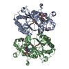
| ||||||||||||||||||||||||
|---|---|---|---|---|---|---|---|---|---|---|---|---|---|---|---|---|---|---|---|---|---|---|---|---|---|
| 1 | 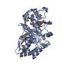
| ||||||||||||||||||||||||
| 2 | 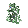
| ||||||||||||||||||||||||
| 単位格子 |
| ||||||||||||||||||||||||
| 非結晶学的対称性 (NCS) | NCSドメイン:
NCSドメイン領域: Component-ID: 1 / Ens-ID: 1 / Beg auth comp-ID: VAL / Beg label comp-ID: VAL / End auth comp-ID: TYR / End label comp-ID: TYR / Auth asym-ID: A / Label asym-ID: A / Auth seq-ID: 3 - 313 / Label seq-ID: 3 - 313
NCSアンサンブル: (詳細: Global NCS restraints between domains: 1 2) NCS oper:
|
-要素
| #1: タンパク質 | 分子量: 36302.082 Da / 分子数: 2 / 由来タイプ: 組換発現 由来: (組換発現) Limosilactobacillus fermentum (バクテリア)遺伝子: BUW47_03110, C1Y38_03730, DBX48_02645, GC247_08580, GDZ34_00450, GJA14_02385, LACFE_CDS1279 発現宿主: #2: 化合物 |   分子量: 616.487 Da / 分子数: 2 / 由来タイプ: 天然 / 式: C34H32FeN4O4 / タイプ: SUBJECT OF INVESTIGATION 分子量: 616.487 Da / 分子数: 2 / 由来タイプ: 天然 / 式: C34H32FeN4O4 / タイプ: SUBJECT OF INVESTIGATION#3: 水 | ChemComp-HOH / |  分子量: 18.015 Da / 分子数: 232 / 由来タイプ: 天然 / 式: H2O 分子量: 18.015 Da / 分子数: 232 / 由来タイプ: 天然 / 式: H2O研究の焦点であるリガンドがあるか | Y | Has protein modification | Y | |
|---|
-実験情報
-実験
| 実験 | 手法: X線回折 / 使用した結晶の数: 1 |
|---|
- 試料調製
試料調製
| 結晶 | マシュー密度: 3.07 Å3/Da / 溶媒含有率: 59.98 % |
|---|---|
| 結晶化 | 温度: 295 K / 手法: 蒸気拡散法, シッティングドロップ法 詳細: 1.0 M lithium sulfate, 0.1 M sodium citrate (pH 5.6) and 0.5 M Ammonium sulfate |
-データ収集
| 回折 | 平均測定温度: 100 K / Serial crystal experiment: N |
|---|---|
| 放射光源 | 由来: シンクロトロン / サイト: SSRF  / ビームライン: BL02U1 / 波長: 0.9792 Å / ビームライン: BL02U1 / 波長: 0.9792 Å |
| 検出器 | タイプ: DECTRIS PILATUS 6M / 検出器: PIXEL / 日付: 2021年12月10日 |
| 放射 | プロトコル: SINGLE WAVELENGTH / 単色(M)・ラウエ(L): M / 散乱光タイプ: x-ray |
| 放射波長 | 波長: 0.9792 Å / 相対比: 1 |
| 反射 | 解像度: 1.83→67.59 Å / Num. obs: 36030 / % possible obs: 92.5 % / 冗長度: 17.6 % / CC1/2: 0.98 / Rmerge(I) obs: 0.163 / Rpim(I) all: 0.04 / Rrim(I) all: 0.168 / Χ2: 0.97 / Net I/σ(I): 14.9 |
| 反射 シェル | 解像度: 1.83→1.87 Å / 冗長度: 12.3 % / Rmerge(I) obs: 2.304 / Mean I/σ(I) obs: 1.3 / Num. unique obs: 687 / CC1/2: 0.372 / Rpim(I) all: 0.655 / Rrim(I) all: 2.41 / Χ2: 0.77 / % possible all: 58.2 |
- 解析
解析
| ソフトウェア |
| |||||||||||||||||||||||||||||||||||||||||||||||||||||||||||||||||||||||||||||||||||||||||||||||||||||||||||||||||||||||||||||||||||||||||||||||||||||||||||||||||||||||||||||||||||||||||||||
|---|---|---|---|---|---|---|---|---|---|---|---|---|---|---|---|---|---|---|---|---|---|---|---|---|---|---|---|---|---|---|---|---|---|---|---|---|---|---|---|---|---|---|---|---|---|---|---|---|---|---|---|---|---|---|---|---|---|---|---|---|---|---|---|---|---|---|---|---|---|---|---|---|---|---|---|---|---|---|---|---|---|---|---|---|---|---|---|---|---|---|---|---|---|---|---|---|---|---|---|---|---|---|---|---|---|---|---|---|---|---|---|---|---|---|---|---|---|---|---|---|---|---|---|---|---|---|---|---|---|---|---|---|---|---|---|---|---|---|---|---|---|---|---|---|---|---|---|---|---|---|---|---|---|---|---|---|---|---|---|---|---|---|---|---|---|---|---|---|---|---|---|---|---|---|---|---|---|---|---|---|---|---|---|---|---|---|---|---|---|---|
| 精密化 | 構造決定の手法: 多重同系置換 / 解像度: 1.83→67.589 Å / Cor.coef. Fo:Fc: 0.947 / Cor.coef. Fo:Fc free: 0.94 / WRfactor Rfree: 0.215 / WRfactor Rwork: 0.184 / SU B: 2.794 / SU ML: 0.078 / Average fsc free: 0 / Average fsc work: 0 / 交差検証法: NONE / ESU R: 0.31 / ESU R Free: 0.225 詳細: Hydrogens have been added in their riding positions. The model represents an ordered domain of a partially disordered crystal. Reflections L=2N+1 were severely affected by crystal disorder ...詳細: Hydrogens have been added in their riding positions. The model represents an ordered domain of a partially disordered crystal. Reflections L=2N+1 were severely affected by crystal disorder and could not be measured. Reflections L=2N were not affected by crystal disorder and were used for structure solution and refinement. Scaling and merging was done in point group 6 2 2, c=42.0A. Refinement was conducted against 50% complete P 3 2 1 data set, c=84A, see associated manuscript for details.
| |||||||||||||||||||||||||||||||||||||||||||||||||||||||||||||||||||||||||||||||||||||||||||||||||||||||||||||||||||||||||||||||||||||||||||||||||||||||||||||||||||||||||||||||||||||||||||||
| 溶媒の処理 | イオンプローブ半径: 0.8 Å / 減衰半径: 0.8 Å / VDWプローブ半径: 1.2 Å / 溶媒モデル: MASK BULK SOLVENT | |||||||||||||||||||||||||||||||||||||||||||||||||||||||||||||||||||||||||||||||||||||||||||||||||||||||||||||||||||||||||||||||||||||||||||||||||||||||||||||||||||||||||||||||||||||||||||||
| 原子変位パラメータ | Biso mean: 22.268 Å2
| |||||||||||||||||||||||||||||||||||||||||||||||||||||||||||||||||||||||||||||||||||||||||||||||||||||||||||||||||||||||||||||||||||||||||||||||||||||||||||||||||||||||||||||||||||||||||||||
| 精密化ステップ | サイクル: LAST / 解像度: 1.83→67.589 Å
| |||||||||||||||||||||||||||||||||||||||||||||||||||||||||||||||||||||||||||||||||||||||||||||||||||||||||||||||||||||||||||||||||||||||||||||||||||||||||||||||||||||||||||||||||||||||||||||
| 拘束条件 |
| |||||||||||||||||||||||||||||||||||||||||||||||||||||||||||||||||||||||||||||||||||||||||||||||||||||||||||||||||||||||||||||||||||||||||||||||||||||||||||||||||||||||||||||||||||||||||||||
| Refine LS restraints NCS | Auth asym-ID: A / Ens-ID: 1 / Refine-ID: X-RAY DIFFRACTION / タイプ: tight positional; tight thermal / Rms dev Biso : 0.10112 Å2 / Rms dev position: 0.00437 Å / Weight Biso : 0.5 / Weight position: 0.05
| |||||||||||||||||||||||||||||||||||||||||||||||||||||||||||||||||||||||||||||||||||||||||||||||||||||||||||||||||||||||||||||||||||||||||||||||||||||||||||||||||||||||||||||||||||||||||||||
| LS精密化 シェル | Refine-ID: X-RAY DIFFRACTION / Total num. of bins used: 20
|
