 Movie
Movie Controller


+ Open data

- Basic information
| Entry | Database: PDB / ID: 7yll | ||||||
|---|---|---|---|---|---|---|---|
| Title | Crystal structure of TTEDbh | ||||||
 Components Components | DNA polymerase IV | ||||||
 Keywords Keywords | TRANSFERASE / polymerase of DinB subfamily / translesion synthesis / DNA binding protein / REPLICATION | ||||||
| Function / homology |  Function and homology information Function and homology informationoxidized DNA binding / DNA synthesis involved in DNA repair / error-prone translesion synthesis / DNA-templated DNA replication / double-stranded DNA binding / DNA-directed DNA polymerase / DNA-directed DNA polymerase activity / magnesium ion binding / cytoplasm Similarity search - Function | ||||||
| Biological species |  Thermoanaerobacter tengcongensis (bacteria) Thermoanaerobacter tengcongensis (bacteria) | ||||||
| Method |  X-RAY DIFFRACTION / SYNCHROTRON / MOLECULAR REPLACEMENT / Resolution: 2.60000921196 Å X-RAY DIFFRACTION / SYNCHROTRON / MOLECULAR REPLACEMENT / Resolution: 2.60000921196 Å | ||||||
 Authors Authors | Yan, X. / Tian, L. / Gao, H. | ||||||
| Funding support | 1items
| ||||||
 Citation Citation | Journal: Int.J.Biol.Macromol. / Year: 2023 Title: Structure and function of extreme TLS DNA polymerase TTEDbh from Thermoanaerobacter tengcongensis. Authors: Tian, L.F. / Gao, H. / Yang, S. / Liu, Y.P. / Li, M. / Xu, W. / Yan, X.X. | ||||||
| History |
|
- Structure visualization
| Structure viewer | Molecule: MolmilJmol/JSmol |
|---|
- Downloads & links
-Download
| PDBx/mmCIF format | 7yll.cif.gz | 92 KB | Display | PDBx/mmCIF format |
|---|---|---|---|---|
| PDB format | pdb7yll.ent.gz | 61.9 KB | Display | PDB format |
| PDBx/mmJSON format | 7yll.json.gz | Tree view | PDBx/mmJSON format | |
| Others |  Other downloads Other downloads |
-Validation report
| Arichive directory | https://data.pdbj.org/pub/pdb/validation_reports/yl/7yllftp://data.pdbj.org/pub/pdb/validation_reports/yl/7yll | HTTPS FTP |
|---|
-Related structure data
| Related structure data |  2rdiS S: Starting model for refinement |
|---|---|
| Similar structure data |
-Links
 PDBj
PDBj

- Assembly
| Deposited unit | 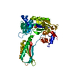
| ||||||||||||
|---|---|---|---|---|---|---|---|---|---|---|---|---|---|
| 1 |
| ||||||||||||
| Unit cell |
| ||||||||||||
| Components on special symmetry positions |
|
-Components
| #1: Protein | Mass: 43988.262 Da / Num. of mol.: 1 Source method: isolated from a genetically manipulated source Source: (gene. exp.) Thermoanaerobacter tengcongensis (strain DSM 15242 / JCM 11007 / NBRC 100824 / MB4) (bacteria)Strain: DSM 15242 / JCM 11007 / NBRC 100824 / MB4 / Gene: dinB, TTE0254 / Production host: |
|---|---|
| #2: Chemical | ChemComp-PO4 /   Mass: 94.971 Da / Num. of mol.: 1 / Source method: obtained synthetically / Formula: PO4 / Feature type: SUBJECT OF INVESTIGATION Mass: 94.971 Da / Num. of mol.: 1 / Source method: obtained synthetically / Formula: PO4 / Feature type: SUBJECT OF INVESTIGATION |
| #3: Chemical | ChemComp-MG /   Mass: 24.305 Da / Num. of mol.: 1 / Source method: obtained synthetically / Formula: Mg / Feature type: SUBJECT OF INVESTIGATION Mass: 24.305 Da / Num. of mol.: 1 / Source method: obtained synthetically / Formula: Mg / Feature type: SUBJECT OF INVESTIGATION |
| #4: Water | ChemComp-HOH /  Mass: 18.015 Da / Num. of mol.: 162 / Source method: isolated from a natural source / Formula: H2O Mass: 18.015 Da / Num. of mol.: 162 / Source method: isolated from a natural source / Formula: H2O |
| Has ligand of interest | Y |
-Experimental details
-Experiment
| Experiment | Method: X-RAY DIFFRACTION / Number of used crystals: 1 |
|---|
- Sample preparation
| Crystal | Density Matthews: 2.33 Å3/Da / Density % sol: 47.29 % |
|---|---|
| Crystal grow | Temperature: 293 K / Method: vapor diffusion, hanging drop / Details: 500 mM NaCl, 5 mM Mg3(PO4)2, 18% [w/v] PEG 3350 |
-Data collection
| Diffraction | Mean temperature: 100 K / Serial crystal experiment: N |
|---|---|
| Diffraction source | Source: SYNCHROTRON / Site: SSRF  / Beamline: BL19U1 / Wavelength: 0.97929 Å / Beamline: BL19U1 / Wavelength: 0.97929 Å |
| Detector | Type: DECTRIS PILATUS 6M / Detector: PIXEL / Date: Jul 18, 2018 |
| Radiation | Protocol: SINGLE WAVELENGTH / Monochromatic (M) / Laue (L): M / Scattering type: x-ray |
| Radiation wavelength | Wavelength: 0.97929 Å / Relative weight: 1 |
| Reflection | Resolution: 2.6→14.88 Å / Num. obs: 22314 / % possible obs: 98.2 % / Redundancy: 7.1 % / Biso Wilson estimate: 46.7017201672 Å2 / CC1/2: 0.98 / Net I/σ(I): 133.9 |
| Reflection shell | Resolution: 2.6→2.77 Å / Num. unique obs: 2185 / CC1/2: 0.998 |
- Processing
| Software |
| ||||||||||||||||||||||||||||||||||||||||||
|---|---|---|---|---|---|---|---|---|---|---|---|---|---|---|---|---|---|---|---|---|---|---|---|---|---|---|---|---|---|---|---|---|---|---|---|---|---|---|---|---|---|---|---|
| Refinement | Method to determine structure: MOLECULAR REPLACEMENT Starting model: 2RDI Resolution: 2.60000921196→14.8769609907 Å / SU ML: 0.310977302148 / Cross valid method: FREE R-VALUE / σ(F): 1.33649243903 / Phase error: 26.6724387516 Stereochemistry target values: GeoStd + Monomer Library + CDL v1.2
| ||||||||||||||||||||||||||||||||||||||||||
| Solvent computation | Shrinkage radii: 0.9 Å / VDW probe radii: 1.11 Å / Solvent model: FLAT BULK SOLVENT MODEL | ||||||||||||||||||||||||||||||||||||||||||
| Displacement parameters | Biso mean: 55.012083344 Å2 | ||||||||||||||||||||||||||||||||||||||||||
| Refinement step | Cycle: LAST / Resolution: 2.60000921196→14.8769609907 Å
| ||||||||||||||||||||||||||||||||||||||||||
| Refine LS restraints |
| ||||||||||||||||||||||||||||||||||||||||||
| LS refinement shell |
|
