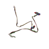
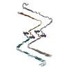
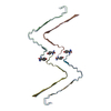
+ Open data
- Basic information
| Entry | Database: PDB / ID: 7yl7 | ||||||||||||
|---|---|---|---|---|---|---|---|---|---|---|---|---|---|
| Title | Structure of hIAPP-TF-type3 | ||||||||||||
 Components Components | Islet amyloid polypeptide | ||||||||||||
 Keywords Keywords | PROTEIN FIBRIL / hIAPP / amylin | ||||||||||||
| Function / homology |  Function and homology information Function and homology informationamylin receptor 3 signaling pathway / amylin receptor 2 signaling pathway / amylin receptor 1 signaling pathway / amylin receptor signaling pathway / Calcitonin-like ligand receptors / negative regulation of amyloid fibril formation / negative regulation of bone resorption / eating behavior / negative regulation of osteoclast differentiation / Regulation of gene expression in beta cells ...amylin receptor 3 signaling pathway / amylin receptor 2 signaling pathway / amylin receptor 1 signaling pathway / amylin receptor signaling pathway / Calcitonin-like ligand receptors / negative regulation of amyloid fibril formation / negative regulation of bone resorption / eating behavior / negative regulation of osteoclast differentiation / Regulation of gene expression in beta cells / positive regulation of cAMP/PKA signal transduction / bone resorption / negative regulation of protein-containing complex assembly / sensory perception of pain / positive regulation of calcium-mediated signaling / osteoclast differentiation / hormone activity / cell-cell signaling / amyloid-beta binding / G alpha (s) signalling events / positive regulation of MAPK cascade / positive regulation of apoptotic process / receptor ligand activity / Amyloid fiber formation / signaling receptor binding / neuronal cell body / apoptotic process / lipid binding / signal transduction / extracellular space / extracellular region / identical protein binding Similarity search - Function | ||||||||||||
| Biological species |  Homo sapiens (human) Homo sapiens (human) | ||||||||||||
| Method | ELECTRON MICROSCOPY / helical reconstruction / Resolution: 3.3 Å | ||||||||||||
 Authors Authors | Li, D. / Zhang, X. | ||||||||||||
| Funding support |  China, 3items China, 3items
| ||||||||||||
 Citation Citation |  Journal: iScience / Year: 2022 Journal: iScience / Year: 2022Title: A new polymorphism of human amylin fibrils with similar protofilaments and a conserved core. Authors: Dongyu Li / Xueli Zhang / Youwang Wang / Haonan Zhang / Kai Song / Keyan Bao / Ping Zhu /  Abstract: Pancreatic amyloid deposits composed of a fibrillar form of the human islet amyloid polypeptide (hIAPP) are the pathological hallmark of type 2 diabetes (T2D). Although various cryo-EM structures of ...Pancreatic amyloid deposits composed of a fibrillar form of the human islet amyloid polypeptide (hIAPP) are the pathological hallmark of type 2 diabetes (T2D). Although various cryo-EM structures of polymorphic hIAPP fibrils were reported, the underlying polymorphic mechanism of hIAPP remains elusive. Meanwhile, the structure of hIAPP fibrils with all residues visible in the fibril core is not available. Here, we report the full-length structures of two different polymorphs of hIAPP fibrils, namely slim form (SF, dimer) and thick form (TF, tetramer), formed in a salt-free environment, which share a similar ζ-shaped protofilament but differ in inter-protofilament interfaces. In the absence of salt, electrostatic interactions were found to play a dominant role in stabilizing the fibril structure, suggesting an antagonistic effect between electrostatic and hydrophobic interactions in different salt concentrations environments. Our results shed light on understanding the mechanism of amyloid fibril polymorphism. | ||||||||||||
| History |
|
- Structure visualization
| Structure viewer | Molecule:  Molmil Molmil Jmol/JSmol Jmol/JSmol |
|---|
- Downloads & links
- Download
| PDBx/mmCIF format |  7yl7.cif.gz 7yl7.cif.gz | 71.7 KB | Display |  PDBx/mmCIF format PDBx/mmCIF format |
|---|---|---|---|---|
| PDB format |  pdb7yl7.ent.gz pdb7yl7.ent.gz | 56.7 KB | Display |  PDB format PDB format |
| PDBx/mmJSON format |  7yl7.json.gz 7yl7.json.gz | Tree view |  PDBx/mmJSON format PDBx/mmJSON format | |
| Others |  Other downloads Other downloads |
-Validation report
| Summary document |  7yl7_validation.pdf.gz 7yl7_validation.pdf.gz | 1 MB | Display |  wwPDB validaton report wwPDB validaton report |
|---|---|---|---|---|
| Full document |  7yl7_full_validation.pdf.gz 7yl7_full_validation.pdf.gz | 1 MB | Display | |
| Data in XML |  7yl7_validation.xml.gz 7yl7_validation.xml.gz | 28.4 KB | Display | |
| Data in CIF |  7yl7_validation.cif.gz 7yl7_validation.cif.gz | 40.2 KB | Display | |
| Arichive directory |  https://data.pdbj.org/pub/pdb/validation_reports/yl/7yl7 https://data.pdbj.org/pub/pdb/validation_reports/yl/7yl7 ftp://data.pdbj.org/pub/pdb/validation_reports/yl/7yl7 ftp://data.pdbj.org/pub/pdb/validation_reports/yl/7yl7 | HTTPS FTP |
-Related structure data
| Related structure data |  33903MC  7ykwC  7yl0C  7yl3C M: map data used to model this data C: citing same article ( |
|---|---|
| Similar structure data | Similarity search - Function & homology  F&H Search F&H Search |
- Links
Links
- Assembly
| Deposited unit | 
|
|---|---|
| 1 |
|
- Components
| #1: Protein/peptide | Mass: 3908.319 Da / Num. of mol.: 12 / Source method: obtained synthetically / Source: (synth.)  Homo sapiens (human) / References: UniProt: P10997 Homo sapiens (human) / References: UniProt: P10997Has ligand of interest | N | |
|---|
-Experimental details
-Experiment
| Experiment | Method: ELECTRON MICROSCOPY |
|---|---|
| EM experiment | Aggregation state: HELICAL ARRAY / 3D reconstruction method: helical reconstruction |
- Sample preparation
| Component | Name: no / Type: ORGANELLE OR CELLULAR COMPONENT / Entity ID: all / Source: SYNTHETIC |
|---|---|
| Source (natural) | Organism:  Homo sapiens (human) Homo sapiens (human) |
| Buffer solution | pH: 7 |
| Specimen | Embedding applied: NO / Shadowing applied: NO / Staining applied: NO / Vitrification applied: NO |
- Electron microscopy imaging
| Experimental equipment |  Model: Titan Krios / Image courtesy: FEI Company |
|---|---|
| Microscopy | Model: FEI TITAN KRIOS |
| Electron gun | Electron source:  FIELD EMISSION GUN / Accelerating voltage: 300 kV / Illumination mode: FLOOD BEAM FIELD EMISSION GUN / Accelerating voltage: 300 kV / Illumination mode: FLOOD BEAM |
| Electron lens | Mode: BRIGHT FIELD / Nominal defocus max: 1500 nm / Nominal defocus min: 1000 nm / Cs: 2.8 mm |
| Image recording | Average exposure time: 3 sec. / Electron dose: 60 e/Å2 / Detector mode: SUPER-RESOLUTION / Film or detector model: GATAN K2 SUMMIT (4k x 4k) |
| Image scans | Movie frames/image: 32 |
- Processing
| Software | Name: PHENIX / Version: 1.19.2_4158: / Classification: refinement | ||||||||||||||||||||||||
|---|---|---|---|---|---|---|---|---|---|---|---|---|---|---|---|---|---|---|---|---|---|---|---|---|---|
| EM software |
| ||||||||||||||||||||||||
| CTF correction | Type: NONE | ||||||||||||||||||||||||
| Helical symmerty | Angular rotation/subunit: 179.41 ° / Axial rise/subunit: 2.35 Å / Axial symmetry: C1 | ||||||||||||||||||||||||
| 3D reconstruction | Resolution: 3.3 Å / Resolution method: FSC 0.143 CUT-OFF / Num. of particles: 29587 / Symmetry type: HELICAL | ||||||||||||||||||||||||
| Refine LS restraints |
|
 Movie
Movie Controller


 PDBj
PDBj


