- PDB-7yke: Crystal structure of chondroitin ABC lyase I in complex with chon... -
+
Open data
ID or keywords:
Loading...
-
Basic information
Entry
Database: PDB / ID: 7yke
Title
Crystal structure of chondroitin ABC lyase I in complex with chondroitin disaccharide 4,6-sulfate
Components
Chondroitin sulfate ABC endolyase
Keywords
LYASE / Complex / Polysaccharide lyase family 8 / Carbohydrate-binding
Function / homology
Function and homology information
chondroitin-sulfate-ABC endolyase / chondroitin-sulfate-ABC endolyase activity / glycosaminoglycan catabolic process / carbohydrate binding / carbohydrate metabolic process / periplasmic space / extracellular region / metal ion binding Similarity search - Function
Lyase, N-terminal / Lyase, catalytic / Chondroitin sulfate ABC endolyase / Chondroitin sulfate ABC lyase / Lyase, N terminal / Lyase, catalytic / Polysaccharide lyase family 8, C-terminal / Polysaccharide lyase family 8, C-terminal beta-sandwich domain / Polysaccharide lyase family 8, central domain / Polysaccharide lyase family 8, super-sandwich domain ...Lyase, N-terminal / Lyase, catalytic / Chondroitin sulfate ABC endolyase / Chondroitin sulfate ABC lyase / Lyase, N terminal / Lyase, catalytic / Polysaccharide lyase family 8, C-terminal / Polysaccharide lyase family 8, C-terminal beta-sandwich domain / Polysaccharide lyase family 8, central domain / Polysaccharide lyase family 8, super-sandwich domain / Polysaccharide lyase family 8-like, C-terminal / Chondroitin AC/alginate lyase / Glycoside hydrolase-type carbohydrate-binding / Galactose mutarotase-like domain superfamily / Galactose-binding-like domain superfamily Similarity search - Domain/homology
Resolution: 1.88→49.07 Å / Cor.coef. Fo:Fc: 0.963 / Cor.coef. Fo:Fc free: 0.944 / SU B: 6.421 / SU ML: 0.097 / Cross valid method: THROUGHOUT / σ(F): 0 / ESU R: 0.134 / ESU R Free: 0.13 / Stereochemistry target values: MAXIMUM LIKELIHOOD Details: U VALUES : WITH TLS ADDED HYDROGENS HAVE BEEN ADDED IN THE RIDING POSITIONS
Rfactor
Num. reflection
% reflection
Selection details
Rfree
0.2127
4380
5 %
RANDOM
Rwork
0.1692
-
-
-
obs
0.1714
83515
99.96 %
-
Solvent computation
Ion probe radii: 0.8 Å / Shrinkage radii: 0.8 Å / VDW probe radii: 1.2 Å / Solvent model: MASK
In the structure databanks used in Yorodumi, some data are registered as the other names, "COVID-19 virus" and "2019-nCoV". Here are the details of the virus and the list of structure data.
Jan 31, 2019. EMDB accession codes are about to change! (news from PDBe EMDB page)
EMDB accession codes are about to change! (news from PDBe EMDB page)
The allocation of 4 digits for EMDB accession codes will soon come to an end. Whilst these codes will remain in use, new EMDB accession codes will include an additional digit and will expand incrementally as the available range of codes is exhausted. The current 4-digit format prefixed with “EMD-” (i.e. EMD-XXXX) will advance to a 5-digit format (i.e. EMD-XXXXX), and so on. It is currently estimated that the 4-digit codes will be depleted around Spring 2019, at which point the 5-digit format will come into force.
The EM Navigator/Yorodumi systems omit the EMD- prefix.
Related info.:Q: What is EMD? / ID/Accession-code notation in Yorodumi/EM Navigator
Yorodumi is a browser for structure data from EMDB, PDB, SASBDB, etc.
This page is also the successor to EM Navigator detail page, and also detail information page/front-end page for Omokage search.
The word "yorodu" (or yorozu) is an old Japanese word meaning "ten thousand". "mi" (miru) is to see.
Related info.:EMDB / PDB / SASBDB / Comparison of 3 databanks / Yorodumi Search / Aug 31, 2016. New EM Navigator & Yorodumi / Yorodumi Papers / Jmol/JSmol / Function and homology information / Changes in new EM Navigator and Yorodumi
 Movie
Movie Controller

 Yorodumi
 Open data
 Basic information
 Components
 Keywords
 Function and homology information
 Proteus vulgaris (bacteria)
 X-RAY DIFFRACTION /
  Authors
 Citation
 Structure visualization
 Downloads & links
 Other downloads
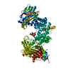
 PDBj
PDBj


 Assembly
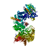

 Mass: 24.305 Da / Num. of mol.: 1 / Source method: obtained synthetically / Formula: Mg
 Mass: 18.015 Da / Num. of mol.: 741 / Source method: isolated from a natural source / Formula: H2O
 Sample preparation
 / Beamline: BL-5A / Wavelength: 1 Å
 Processing