 Movie
Movie Controller


+ Open data

- Basic information
| Entry | Database: PDB / ID: 7yk1 | |||||||||
|---|---|---|---|---|---|---|---|---|---|---|
| Title | Structural basis of human PRPS2 filaments | |||||||||
 Components Components | Ribose-phosphate pyrophosphokinase 2 | |||||||||
 Keywords Keywords | TRANSFERASE / phosphoribosylpyrophosphate synthetase / complex / filament | |||||||||
| Function / homology |  Function and homology information Function and homology information5-Phosphoribose 1-diphosphate biosynthesis / ribonucleoside monophosphate biosynthetic process / ribose-phosphate diphosphokinase / ribose phosphate diphosphokinase activity / 5-phosphoribose 1-diphosphate biosynthetic process / pentose-phosphate shunt / purine nucleotide biosynthetic process / nucleobase-containing compound metabolic process / kinase activity / magnesium ion binding ...5-Phosphoribose 1-diphosphate biosynthesis / ribonucleoside monophosphate biosynthetic process / ribose-phosphate diphosphokinase / ribose phosphate diphosphokinase activity / 5-phosphoribose 1-diphosphate biosynthetic process / pentose-phosphate shunt / purine nucleotide biosynthetic process / nucleobase-containing compound metabolic process / kinase activity / magnesium ion binding / protein homodimerization activity / ATP binding / identical protein binding / cytosol / cytoplasm Similarity search - Function | |||||||||
| Biological species |  Homo sapiens (human) Homo sapiens (human) | |||||||||
| Method | ELECTRON MICROSCOPY / single particle reconstruction / cryo EM / Resolution: 3.08 Å | |||||||||
 Authors Authors | Lu, G.M. / Hu, H.H. / Liu, J.L. | |||||||||
| Funding support |  China, 2items China, 2items
| |||||||||
 Citation Citation | Journal: Cell Biosci / Year: 2023 Title: Structural basis of human PRPS2 filaments. Authors: Guang-Ming Lu / Huan-Huan Hu / Chia-Chun Chang / Jiale Zhong / Xian Zhou / Chen-Jun Guo / Tianyi Zhang / Yi-Lan Li / Boqi Yin / Ji-Long Liu /  Abstract: BACKGROUND: PRPP synthase (PRPS) transfers the pyrophosphate groups from ATP to ribose-5-phosphate to produce 5-phosphate ribose-1-pyrophosphate (PRPP), a key intermediate in the biosynthesis of ...BACKGROUND: PRPP synthase (PRPS) transfers the pyrophosphate groups from ATP to ribose-5-phosphate to produce 5-phosphate ribose-1-pyrophosphate (PRPP), a key intermediate in the biosynthesis of several metabolites including nucleotides, dinucleotides and some amino acids. There are three PRPS isoforms encoded in human genome. While human PRPS1 (hPRPS1) and human PRPS2 (hPRPS2) are expressed in most tissues, human PRPS3 (hPRPS3) is exclusively expressed in testis. Although hPRPS1 and hPRPS2 share 95% sequence identity, hPRPS2 has been shown to be less sensitive to allosteric inhibition and specifically upregulated in certain cancers in the translational level. Recent studies demonstrate that PRPS can form a subcellular compartment termed the cytoophidium in multiple organisms across prokaryotes and eukaryotes. Forming filaments and cytoophidia is considered as a distinctive mechanism involving the polymerization of the protein. Previously we solved the filament structures of Escherichia coli PRPS (ecPRPS) using cryo-electron microscopy (cryo-EM) . RESULTS: Order to investigate the function and molecular mechanism of hPRPS2 polymerization, here we solve the polymer structure of hPRPS2 at 3.08 Å resolution. hPRPS2 hexamers stack into polymers ...RESULTS: Order to investigate the function and molecular mechanism of hPRPS2 polymerization, here we solve the polymer structure of hPRPS2 at 3.08 Å resolution. hPRPS2 hexamers stack into polymers in the conditions with the allosteric/competitive inhibitor ADP. The binding modes of ADP at the canonical allosteric site and at the catalytic active site are clearly determined. A point mutation disrupting the inter-hexamer interaction prevents hPRPS2 polymerization and results in significantly reduced catalytic activity. CONCLUSION: Findings suggest that the regulation of hPRPS2 polymer is distinct from ecPRPS polymer and provide new insights to the regulation of hPRPS2 with structural basis. | |||||||||
| History |
|
- Structure visualization
| Structure viewer | Molecule: MolmilJmol/JSmol |
|---|
- Downloads & links
-Download
| PDBx/mmCIF format | 7yk1.cif.gz | 434.2 KB | Display | PDBx/mmCIF format |
|---|---|---|---|---|
| PDB format | pdb7yk1.ent.gz | 285.5 KB | Display | PDB format |
| PDBx/mmJSON format | 7yk1.json.gz | Tree view | PDBx/mmJSON format | |
| Others |  Other downloads Other downloads |
-Validation report
| Summary document | 7yk1_validation.pdf.gz | 2 MB | Display | wwPDB validaton report |
|---|---|---|---|---|
| Full document | 7yk1_full_validation.pdf.gz | 2 MB | Display | |
| Data in XML | 7yk1_validation.xml.gz | 63.3 KB | Display | |
| Data in CIF | 7yk1_validation.cif.gz | 90.7 KB | Display | |
| Arichive directory | https://data.pdbj.org/pub/pdb/validation_reports/yk/7yk1ftp://data.pdbj.org/pub/pdb/validation_reports/yk/7yk1 | HTTPS FTP |
-Related structure data
| Related structure data |  33883MC M: map data used to model this data C: citing same article ( |
|---|---|
| Similar structure data |
-Links
 PDBj
PDBj
- Assembly
| Deposited unit | 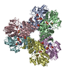
|
|---|---|
| 1 |
|
-Components
| #1: Protein | Mass: 35640.969 Da / Num. of mol.: 6 Source method: isolated from a genetically manipulated source Source: (gene. exp.) Homo sapiens (human) / Gene: PRPS2Production host:  References: UniProt: P11908, ribose-phosphate diphosphokinase #2: Chemical | ChemComp-PO4 /   Mass: 94.971 Da / Num. of mol.: 6 / Source method: obtained synthetically / Formula: PO4 / Feature type: SUBJECT OF INVESTIGATION Mass: 94.971 Da / Num. of mol.: 6 / Source method: obtained synthetically / Formula: PO4 / Feature type: SUBJECT OF INVESTIGATION#3: Chemical | ChemComp-MG /   Mass: 24.305 Da / Num. of mol.: 6 / Source method: obtained synthetically / Formula: Mg / Feature type: SUBJECT OF INVESTIGATION Mass: 24.305 Da / Num. of mol.: 6 / Source method: obtained synthetically / Formula: Mg / Feature type: SUBJECT OF INVESTIGATION#4: Chemical | ChemComp-ADP /   Mass: 427.201 Da / Num. of mol.: 12 / Source method: obtained synthetically / Formula: C10H15N5O10P2 / Feature type: SUBJECT OF INVESTIGATION / Comment: ADP, energy-carrying molecule*YM Mass: 427.201 Da / Num. of mol.: 12 / Source method: obtained synthetically / Formula: C10H15N5O10P2 / Feature type: SUBJECT OF INVESTIGATION / Comment: ADP, energy-carrying molecule*YMHas ligand of interest | Y | |
|---|
-Experimental details
-Experiment
| Experiment | Method: ELECTRON MICROSCOPY |
|---|---|
| EM experiment | Aggregation state: FILAMENT / 3D reconstruction method: single particle reconstruction |
- Sample preparation
| Component | Name: Filament structure of human phosphoribosylpyrophosphate synthetase2. Type: COMPLEX / Entity ID: #1 / Source: RECOMBINANT | |||||||||||||||||||||||||
|---|---|---|---|---|---|---|---|---|---|---|---|---|---|---|---|---|---|---|---|---|---|---|---|---|---|---|
| Molecular weight | Experimental value: NO | |||||||||||||||||||||||||
| Source (natural) | Organism: Homo sapiens (human) | |||||||||||||||||||||||||
| Source (recombinant) | Organism: | |||||||||||||||||||||||||
| Buffer solution | pH: 7.5 | |||||||||||||||||||||||||
| Buffer component |
| |||||||||||||||||||||||||
| Specimen | Embedding applied: NO / Shadowing applied: NO / Staining applied: NO / Vitrification applied: YES | |||||||||||||||||||||||||
| Specimen support | Grid material: GOLD / Grid type: UltrAuFoil R1.2/1.3 | |||||||||||||||||||||||||
| Vitrification | Cryogen name: ETHANE / Humidity: 100 % / Chamber temperature: 277.15 K |
- Electron microscopy imaging
| Experimental equipment |  Model: Titan Krios / Image courtesy: FEI Company |
|---|---|
| Microscopy | Model: FEI TITAN KRIOS |
| Electron gun | Electron source:  FIELD EMISSION GUN / Accelerating voltage: 300 kV / Illumination mode: FLOOD BEAM FIELD EMISSION GUN / Accelerating voltage: 300 kV / Illumination mode: FLOOD BEAM |
| Electron lens | Mode: BRIGHT FIELD / Nominal magnification: 22500 X / Nominal defocus max: 2500 nm / Nominal defocus min: 1000 nm / Cs: 2.7 mm / Alignment procedure: COMA FREE |
| Specimen holder | Cryogen: NITROGEN |
| Image recording | Average exposure time: 4 sec. / Electron dose: 60 e/Å2 / Film or detector model: GATAN K3 BIOQUANTUM (6k x 4k) / Num. of grids imaged: 1 / Num. of real images: 2403 |
- Processing
| Software |
| ||||||||||||||||||||||||
|---|---|---|---|---|---|---|---|---|---|---|---|---|---|---|---|---|---|---|---|---|---|---|---|---|---|
| EM software | Name: RELION / Version: 3.1_beta / Category: particle selection | ||||||||||||||||||||||||
| CTF correction | Type: PHASE FLIPPING AND AMPLITUDE CORRECTION | ||||||||||||||||||||||||
| Particle selection | Num. of particles selected: 681672 | ||||||||||||||||||||||||
| 3D reconstruction | Resolution: 3.08 Å / Resolution method: FSC 0.143 CUT-OFF / Num. of particles: 140303 / Algorithm: FOURIER SPACE / Num. of class averages: 1 / Symmetry type: POINT | ||||||||||||||||||||||||
| Refinement | Cross valid method: NONE Stereochemistry target values: GeoStd + Monomer Library + CDL v1.2 | ||||||||||||||||||||||||
| Displacement parameters | Biso mean: 18.23 Å2 | ||||||||||||||||||||||||
| Refine LS restraints |
|
