| Entry | Database: PDB / ID: 7yjw
|
|---|
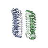
| Title | Structure of Leptospira santarosai serovar shermani LRR protein LSS01692 |
|---|
 Components Components | Membrane protein |
|---|
 Keywords Keywords | MEMBRANE PROTEIN / Leptospira / Leptospirosis / Leucine-rich repeat / CELL ADHESION |
|---|
| Function / homology |  Function and homology information Function and homology information
: / Leucine rich repeat 4 / Leucine Rich repeats (2 copies) / Leucine Rich Repeat / Leucine-rich repeats, bacterial type / Leucine-rich repeat, SDS22-like subfamily / Leucine rich repeat / Leucine-rich repeat, typical subtype / Leucine-rich repeats, typical (most populated) subfamily / Leucine-rich repeat profile. ...: / Leucine rich repeat 4 / Leucine Rich repeats (2 copies) / Leucine Rich Repeat / Leucine-rich repeats, bacterial type / Leucine-rich repeat, SDS22-like subfamily / Leucine rich repeat / Leucine-rich repeat, typical subtype / Leucine-rich repeats, typical (most populated) subfamily / Leucine-rich repeat profile. / Leucine-rich repeat / Leucine-rich repeat domain superfamilySimilarity search - Domain/homology |
|---|
| Biological species |  Leptospira santarosai serovar Shermani (bacteria) Leptospira santarosai serovar Shermani (bacteria) |
|---|
| Method |  X-RAY DIFFRACTION / SYNCHROTRON / MOLECULAR REPLACEMENT / Resolution: 3.2 Å X-RAY DIFFRACTION / SYNCHROTRON / MOLECULAR REPLACEMENT / Resolution: 3.2 Å |
|---|
 Authors Authors | Wu, C.T. / Hsu, S.H. / Yang, C.W. / Sun, Y.J. |
|---|
| Funding support |  Taiwan, 1items Taiwan, 1items | Organization | Grant number | Country |
|---|
| Ministry of Science and Technology (MoST, Taiwan) | | Taiwan |
|
|---|
 Citation Citation | Journal: Febs J. / Year: 2023
Title: Crystal structure of Leptospira LSS_01692 reveals a dimeric structure and induces inflammatory responses through Toll-like receptor 2-dependent NF-kappa B and MAPK signal transduction pathways.
Authors: Hsu, S.H. / Wu, C.T. / Sun, Y.J. / Chang, M.Y. / Li, C. / Ko, Y.C. / Chou, L.F. / Yang, C.W. |
|---|
| History | | Deposition | Jul 20, 2022 | Deposition site: PDBJ / Processing site: PDBJ |
|---|
| Revision 1.0 | Jun 28, 2023 | Provider: repository / Type: Initial release |
|---|
| Revision 1.1 | Oct 4, 2023 | Group: Data collection / Database references
Category: chem_comp_atom / chem_comp_bond ...chem_comp_atom / chem_comp_bond / citation / citation_author
Item: _citation.journal_volume / _citation.page_first ..._citation.journal_volume / _citation.page_first / _citation.page_last / _citation.title / _citation_author.identifier_ORCID |
|---|
| Revision 1.2 | Nov 29, 2023 | Group: Refinement description / Category: pdbx_initial_refinement_model |
|---|
|
|---|
 Movie
Movie Controller

 Yorodumi
 Open data
 Basic information
 Structure visualization
 Downloads & links
 Other downloads
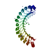
 PDBj
PDBj


 Assembly