 Movie
Movie Controller

[English] 日本語
 Yorodumi
- PDB-7yjl: Melbournevirus major capsid protein built from 4.4A reconstruction -
+ Open data

- Basic information
| Entry | Database: PDB / ID: 7yjl | |||||||||||||||
|---|---|---|---|---|---|---|---|---|---|---|---|---|---|---|---|---|
| Title | Melbournevirus major capsid protein built from 4.4A reconstruction | |||||||||||||||
 Components Components | Major capsid protein | |||||||||||||||
 Keywords Keywords | VIRAL PROTEIN / MCP / Melbournevirus / giant virus / major capsid protein | |||||||||||||||
| Function / homology | Major capsid protein, N-terminal / Major capsid protein N-terminus / Major capsid protein, C-terminal / Major capsid protein, C-terminal domain superfamily / Large eukaryotic DNA virus major capsid protein / Group II dsDNA virus coat/capsid protein / viral capsid / structural molecule activity / Major capsid protein Function and homology information Function and homology information | |||||||||||||||
| Biological species |  Melbournevirus Melbournevirus | |||||||||||||||
| Method | ELECTRON MICROSCOPY / single particle reconstruction / cryo EM / Resolution: 4.42 Å | |||||||||||||||
 Authors Authors | Burton-Smith, R.N. / Okamoto, K. / Murata, K. | |||||||||||||||
| Funding support |  Japan, Japan,  Sweden, 4items Sweden, 4items
| |||||||||||||||
 Citation Citation | Journal: To Be Published Title: The 4.4A structure of the giant Melbournevirus virion belonging to the Marseilleviridae family Authors: Burton-Smith, R.N. / Reddy, H.K.N. / Svenda, M. / Abergel, C. / Okamoto, K. / Murata, K. | |||||||||||||||
| History |
|
- Structure visualization
| Structure viewer | Molecule: MolmilJmol/JSmol |
|---|
- Downloads & links
-Download
| PDBx/mmCIF format | 7yjl.cif.gz | 157.4 KB | Display | PDBx/mmCIF format |
|---|---|---|---|---|
| PDB format | pdb7yjl.ent.gz | 124.6 KB | Display | PDB format |
| PDBx/mmJSON format | 7yjl.json.gz | Tree view | PDBx/mmJSON format | |
| Others |  Other downloads Other downloads |
-Validation report
| Arichive directory | https://data.pdbj.org/pub/pdb/validation_reports/yj/7yjlftp://data.pdbj.org/pub/pdb/validation_reports/yj/7yjl | HTTPS FTP |
|---|
-Related structure data
| Related structure data |  31529MC M: map data used to model this data C: citing same article ( |
|---|---|
| Similar structure data |


-Links
 PDBj
PDBj

- Assembly
| Deposited unit | 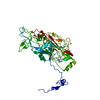
|
|---|---|
| 1 |
|
-Components
| #1: Protein | Mass: 50896.844 Da / Num. of mol.: 1 / Source method: isolated from a natural source / Source: (natural) Melbournevirus / References: UniProt: A0A097I267 |
|---|---|
| Has protein modification | N |
-Experimental details
-Experiment
| Experiment | Method: ELECTRON MICROSCOPY |
|---|---|
| EM experiment | Aggregation state: PARTICLE / 3D reconstruction method: single particle reconstruction |
- Sample preparation
| Component | Name: Melbournevirus / Type: VIRUS / Entity ID: all / Source: NATURAL |
|---|---|
| Source (natural) | Organism: Melbournevirus |
| Details of virus | Empty: NO / Enveloped: NO / Isolate: STRAIN / Type: VIRION |
| Buffer solution | pH: 7.4 |
| Specimen | Embedding applied: NO / Shadowing applied: NO / Staining applied: NO / Vitrification applied: YES |
| Vitrification | Cryogen name: ETHANE |
- Electron microscopy imaging
| Experimental equipment |  Model: Titan Krios / Image courtesy: FEI Company |
|---|---|
| Microscopy | Model: FEI TITAN KRIOS |
| Electron gun | Electron source:  FIELD EMISSION GUN / Accelerating voltage: 300 kV / Illumination mode: FLOOD BEAM FIELD EMISSION GUN / Accelerating voltage: 300 kV / Illumination mode: FLOOD BEAM |
| Electron lens | Mode: BRIGHT FIELD / Nominal defocus max: 3000 nm / Nominal defocus min: 2000 nm / Cs: 2.7 mm |
| Image recording | Electron dose: 26.4 e/Å2 / Film or detector model: GATAN K2 SUMMIT (4k x 4k) |
- Processing
| Software | Name: PHENIX / Version: 1.15.2_3472: / Classification: refinement | ||||||||||||||||||||||||
|---|---|---|---|---|---|---|---|---|---|---|---|---|---|---|---|---|---|---|---|---|---|---|---|---|---|
| EM software | Name: PHENIX / Category: model refinement | ||||||||||||||||||||||||
| CTF correction | Type: PHASE FLIPPING AND AMPLITUDE CORRECTION | ||||||||||||||||||||||||
| 3D reconstruction | Resolution: 4.42 Å / Resolution method: FSC 0.143 CUT-OFF / Num. of particles: 187440 / Symmetry type: POINT | ||||||||||||||||||||||||
| Refine LS restraints |
|
