 Movie
Movie Controller

[English] 日本語
 Yorodumi
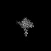
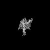
- PDB-7yho: CryoEM structure of Arabidopsis ROS1 in complex with TG mismatch ... -
+ Open data

- Basic information
| Entry | Database: PDB / ID: 7yho | |||||||||||||||||||||||||||||||||||||||||||||
|---|---|---|---|---|---|---|---|---|---|---|---|---|---|---|---|---|---|---|---|---|---|---|---|---|---|---|---|---|---|---|---|---|---|---|---|---|---|---|---|---|---|---|---|---|---|---|
| Title | CryoEM structure of Arabidopsis ROS1 in complex with TG mismatch dsDNA at 3.3 Angstroms resolution | |||||||||||||||||||||||||||||||||||||||||||||
 Components Components |
| |||||||||||||||||||||||||||||||||||||||||||||
 Keywords Keywords | DNA BINDING PROTEIN/DNA / ROS1 / DNA glycosylase / DNA demethylation / DNA BINDING PROTEIN / DNA BINDING PROTEIN-DNA complex | |||||||||||||||||||||||||||||||||||||||||||||
| Function / homology |  Function and homology information Function and homology informationDNA demethylase activity / chromosomal 5-methylcytosine DNA demethylation pathway / positive regulation of male gonad development / Transcriptional regulation of testis differentiation / sex differentiation / DNA N-glycosylase activity / male sex determination / defense response to fungus / DNA-(apurinic or apyrimidinic site) endonuclease activity / class I DNA-(apurinic or apyrimidinic site) endonuclease activity ...DNA demethylase activity / chromosomal 5-methylcytosine DNA demethylation pathway / positive regulation of male gonad development / Transcriptional regulation of testis differentiation / sex differentiation / DNA N-glycosylase activity / male sex determination / defense response to fungus / DNA-(apurinic or apyrimidinic site) endonuclease activity / class I DNA-(apurinic or apyrimidinic site) endonuclease activity / DNA-(apurinic or apyrimidinic site) lyase / Deactivation of the beta-catenin transactivating complex / base-excision repair / 4 iron, 4 sulfur cluster binding / DNA-binding transcription activator activity, RNA polymerase II-specific / DNA-binding transcription factor binding / DNA-binding transcription factor activity, RNA polymerase II-specific / cell differentiation / calmodulin binding / nuclear speck / RNA polymerase II cis-regulatory region sequence-specific DNA binding / DNA repair / positive regulation of gene expression / positive regulation of DNA-templated transcription / chromatin / nucleolus / positive regulation of transcription by RNA polymerase II / DNA binding / nucleoplasm / metal ion binding / nucleus / cytoplasm Similarity search - Function | |||||||||||||||||||||||||||||||||||||||||||||
| Biological species |  Homo sapiens (human) Homo sapiens (human) | |||||||||||||||||||||||||||||||||||||||||||||
| Method | ELECTRON MICROSCOPY / single particle reconstruction / cryo EM / Resolution: 3.3 Å | |||||||||||||||||||||||||||||||||||||||||||||
 Authors Authors | Du, X. / Du, J. | |||||||||||||||||||||||||||||||||||||||||||||
| Funding support | 1items
| |||||||||||||||||||||||||||||||||||||||||||||
 Citation Citation | Journal: Nat Plants / Year: 2023 Title: Molecular basis of the plant ROS1-mediated active DNA demethylation. Authors: Xuan Du / Zhenlin Yang / Guohui Xie / Changshi Wang / Laixing Zhang / Kaige Yan / Maojun Yang / Sisi Li / Jian-Kang Zhu / Jiamu Du /   Abstract: Active DNA demethylation plays a crucial role in eukaryotic gene imprinting and antagonizing DNA methylation. The plant-specific REPRESSOR OF SILENCING 1/DEMETER (ROS1/DME) family of enzymes directly ...Active DNA demethylation plays a crucial role in eukaryotic gene imprinting and antagonizing DNA methylation. The plant-specific REPRESSOR OF SILENCING 1/DEMETER (ROS1/DME) family of enzymes directly excise 5-methyl-cytosine (5mC), representing an efficient DNA demethylation pathway distinct from that of animals. Here, we report the cryo-electron microscopy structures of an Arabidopsis ROS1 catalytic fragment in complex with substrate DNA, mismatch DNA and reaction intermediate, respectively. The substrate 5mC is flipped-out from the DNA duplex and subsequently recognized by the ROS1 base-binding pocket through hydrophobic and hydrogen-bonding interactions towards the 5-methyl group and Watson-Crick edge respectively, while the different protonation states of the bases determine the substrate preference for 5mC over T:G mismatch. Together with the structure of the reaction intermediate complex, our structural and biochemical studies revealed the molecular basis for substrate specificity, as well as the reaction mechanism underlying 5mC demethylation by the ROS1/DME family of plant-specific DNA demethylases. | |||||||||||||||||||||||||||||||||||||||||||||
| History |
|
- Structure visualization
| Structure viewer | Molecule: MolmilJmol/JSmol |
|---|
- Downloads & links
-Download
| PDBx/mmCIF format | 7yho.cif.gz | 114.1 KB | Display | PDBx/mmCIF format |
|---|---|---|---|---|
| PDB format | pdb7yho.ent.gz | 76.1 KB | Display | PDB format |
| PDBx/mmJSON format | 7yho.json.gz | Tree view | PDBx/mmJSON format | |
| Others |  Other downloads Other downloads |
-Validation report
| Arichive directory | https://data.pdbj.org/pub/pdb/validation_reports/yh/7yhoftp://data.pdbj.org/pub/pdb/validation_reports/yh/7yho | HTTPS FTP |
|---|
-Related structure data
| Related structure data | 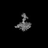 33832MC 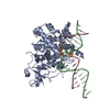 7yhpC  7yhqC M: map data used to model this data C: citing same article ( |
|---|---|
| Similar structure data |
-Links
 PDBj
PDBj


- Assembly
| Deposited unit | 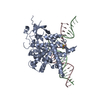
|
|---|---|
| 1 |
|
-Components
| #1: Protein | Mass: 86260.859 Da / Num. of mol.: 1 / Mutation: D971N Source method: isolated from a genetically manipulated source Details: Fusion protein of residues 56 to 130 of database UNP Q05066, Linker, residues 511 to 1393 of database UNP Q9SJQ6, with deletion of residues 665-835 and 1074-1109. Source: (gene. exp.) Homo sapiens (human), (gene. exp.) Gene: SRY, TDF, ROS1, DML1, At2g36490, F1O11.12 Production host:  References: UniProt: Q05066, UniProt: Q9SJQ6, DNA-(apurinic or apyrimidinic site) lyase |
|---|---|
| #2: DNA chain | Mass: 12394.990 Da / Num. of mol.: 1 Source method: isolated from a genetically manipulated source Source: (gene. exp.) |
| #3: DNA chain | Mass: 12244.859 Da / Num. of mol.: 1 Source method: isolated from a genetically manipulated source Source: (gene. exp.) |
| #4: Chemical | ChemComp-SF4 /   Mass: 351.640 Da / Num. of mol.: 1 / Source method: obtained synthetically / Formula: Fe4S4 / Feature type: SUBJECT OF INVESTIGATION Mass: 351.640 Da / Num. of mol.: 1 / Source method: obtained synthetically / Formula: Fe4S4 / Feature type: SUBJECT OF INVESTIGATION |
| Has ligand of interest | Y |
| Has protein modification | N |
-Experimental details
-Experiment
| Experiment | Method: ELECTRON MICROSCOPY |
|---|---|
| EM experiment | Aggregation state: PARTICLE / 3D reconstruction method: single particle reconstruction |
- Sample preparation
| Component |
| ||||||||||||||||||||||||
|---|---|---|---|---|---|---|---|---|---|---|---|---|---|---|---|---|---|---|---|---|---|---|---|---|---|
| Source (natural) |
| ||||||||||||||||||||||||
| Source (recombinant) |
| ||||||||||||||||||||||||
| Buffer solution | pH: 8 | ||||||||||||||||||||||||
| Specimen | Conc.: 0.7 mg/ml / Embedding applied: NO / Shadowing applied: NO / Staining applied: NO / Vitrification applied: YES | ||||||||||||||||||||||||
| Vitrification | Cryogen name: ETHANE / Humidity: 100 % |
- Electron microscopy imaging
| Experimental equipment |  Model: Titan Krios / Image courtesy: FEI Company |
|---|---|
| Microscopy | Model: FEI TITAN KRIOS |
| Electron gun | Electron source: OTHER / Accelerating voltage: 300 kV / Illumination mode: OTHER |
| Electron lens | Mode: OTHER / Nominal defocus max: 2500 nm / Nominal defocus min: 1000 nm |
| Image recording | Average exposure time: 1.3467 sec. / Electron dose: 1.2 e/Å2 / Film or detector model: GATAN K3 (6k x 4k) |
- Processing
| Software | Name: PHENIX / Version: 1.19.2_4158: / Classification: refinement | ||||||||||||||||||||||||
|---|---|---|---|---|---|---|---|---|---|---|---|---|---|---|---|---|---|---|---|---|---|---|---|---|---|
| EM software | Name: PHENIX / Category: model refinement | ||||||||||||||||||||||||
| CTF correction | Type: NONE | ||||||||||||||||||||||||
| 3D reconstruction | Resolution: 3.3 Å / Resolution method: FSC 0.143 CUT-OFF / Num. of particles: 111356 / Symmetry type: POINT | ||||||||||||||||||||||||
| Refine LS restraints |
|
