 Movie
Movie Controller

[English] 日本語
 Yorodumi
- PDB-7yhj: Effector binding domain of LysR-Type transcription factor LrhA fr... -
+ Open data

- Basic information
| Entry | Database: PDB / ID: 7yhj | ||||||
|---|---|---|---|---|---|---|---|
| Title | Effector binding domain of LysR-Type transcription factor LrhA from E. coli | ||||||
 Components Components | Probable HTH-type transcriptional regulator LrhA | ||||||
 Keywords Keywords | TRANSCRIPTION / Transcriptional regulator | ||||||
| Function / homology |  Function and homology information Function and homology informationcis-regulatory region sequence-specific DNA binding / sequence-specific DNA binding / DNA-binding transcription factor activity / negative regulation of DNA-templated transcription / regulation of DNA-templated transcription / DNA-templated transcription / positive regulation of DNA-templated transcription Similarity search - Function | ||||||
| Biological species |  | ||||||
| Method |  X-RAY DIFFRACTION / SYNCHROTRON / MOLECULAR REPLACEMENT / Resolution: 3.237 Å X-RAY DIFFRACTION / SYNCHROTRON / MOLECULAR REPLACEMENT / Resolution: 3.237 Å | ||||||
 Authors Authors | Xie, C. / Jiang, X. | ||||||
| Funding support |  Japan, 1items Japan, 1items
| ||||||
 Citation Citation | Journal: J Mol Biol / Year: 2026 Title: Oligomerization-Dependent Regulation of LrhA Controls Bacterial Flagellar Biosynthesis. Authors: Baichun Niu / Masahide Kikkawa / Xuguang Jiang / Abstract: LysR-type transcriptional regulators (LTTRs) are a diverse family of proteins that regulate various cellular processes, including motility in bacteria. In Escherichia coli, the LTTR LrhA represses ...LysR-type transcriptional regulators (LTTRs) are a diverse family of proteins that regulate various cellular processes, including motility in bacteria. In Escherichia coli, the LTTR LrhA represses flagellar biosynthesis by inhibiting the flhDC operon. However, the structural basis underlying this regulation has remained unclear. Here, we determined both a high-resolution crystal structure and a cryo-EM reconstruction of LrhA, revealing a predominant and stable tetrameric organization with pronounced structural variability in its effector-binding region. Structural and biochemical analyses demonstrate that mutations in these variable regions perturb the oligomeric equilibrium of LrhA, shifting the balance between tetrameric and dimeric species. This shift correlates with enhanced DNA binding affinity and stronger repression of the flhDC promoter. While ligand binding may similarly modulate LrhA activity, our data primarily support a model in which alterations in oligomeric state mediated by the variable regions regulate LrhA function. Together, these findings provide a structural framework for understanding how LrhA controls bacterial motility and offer broader insights into oligomerization-based regulation within the LTTR family. | ||||||
| History |
|
- Structure visualization
| Structure viewer | Molecule: MolmilJmol/JSmol |
|---|
- Downloads & links
-Download
| PDBx/mmCIF format | 7yhj.cif.gz | 306.4 KB | Display | PDBx/mmCIF format |
|---|---|---|---|---|
| PDB format | pdb7yhj.ent.gz | 242.4 KB | Display | PDB format |
| PDBx/mmJSON format | 7yhj.json.gz | Tree view | PDBx/mmJSON format | |
| Others |  Other downloads Other downloads |
-Validation report
| Arichive directory | https://data.pdbj.org/pub/pdb/validation_reports/yh/7yhjftp://data.pdbj.org/pub/pdb/validation_reports/yh/7yhj | HTTPS FTP |
|---|
-Related structure data
| Related structure data |  22xoC  3onmS S: Starting model for refinement C: citing same article ( |
|---|---|
| Similar structure data |

-Links
 PDBj
PDBj- Assembly
| Deposited unit | 
| ||||||||
|---|---|---|---|---|---|---|---|---|---|
| 1 | 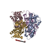
| ||||||||
| 2 | 
| ||||||||
| Unit cell |
|
-Components
| #1: Protein | Mass: 35586.805 Da / Num. of mol.: 8 Source method: isolated from a genetically manipulated source Source: (gene. exp.) #2: Chemical |   Mass: 96.063 Da / Num. of mol.: 2 / Source method: obtained synthetically / Formula: SO4 Mass: 96.063 Da / Num. of mol.: 2 / Source method: obtained synthetically / Formula: SO4#3: Chemical | ChemComp-PEG / |   Mass: 106.120 Da / Num. of mol.: 1 / Source method: obtained synthetically / Formula: C4H10O3 Mass: 106.120 Da / Num. of mol.: 1 / Source method: obtained synthetically / Formula: C4H10O3#4: Water | ChemComp-HOH / |  Mass: 18.015 Da / Num. of mol.: 22 / Source method: isolated from a natural source / Formula: H2O Mass: 18.015 Da / Num. of mol.: 22 / Source method: isolated from a natural source / Formula: H2OHas ligand of interest | N | Has protein modification | N | |
|---|
-Experimental details
-Experiment
| Experiment | Method: X-RAY DIFFRACTION / Number of used crystals: 1 |
|---|
- Sample preparation
| Crystal | Density Matthews: 2.4 Å3/Da / Density % sol: 48.81 % / Description: plate crystal |
|---|---|
| Crystal grow | Temperature: 293 K / Method: vapor diffusion, hanging drop / pH: 7.5 / Details: PEG 3350, 0.1 M HEPES, 0.2 M Ammonium sulfate |
-Data collection
| Diffraction | Mean temperature: 100 K / Serial crystal experiment: N |
|---|---|
| Diffraction source | Source: SYNCHROTRON / Site: SPring-8 / Beamline: BL41XU / Wavelength: 1 Å |
| Detector | Type: DECTRIS EIGER X 16M / Detector: PIXEL / Date: Nov 1, 2018 |
| Radiation | Protocol: SINGLE WAVELENGTH / Monochromatic (M) / Laue (L): M / Scattering type: x-ray |
| Radiation wavelength | Wavelength: 1 Å / Relative weight: 1 |
| Reflection | Resolution: 3.23→50 Å / Num. obs: 43674 / % possible obs: 98.5 % / Redundancy: 5.1 % / CC1/2: 0.977 / Rmerge(I) obs: 0.246 / Rpim(I) all: 0.172 / Rrim(I) all: 0.462 / Net I/σ(I): 3.2 |
| Reflection shell | Resolution: 3.23→3.35 Å / Redundancy: 5.3 % / Rmerge(I) obs: 1.385 / Num. unique obs: 4278 / CC1/2: 0.5 / Rpim(I) all: 0.89 / Rrim(I) all: 2.385 / % possible all: 98.1 |
- Processing
| Software |
| ||||||||||||||||||||||||||||||||||||||||||||||||||||||||||||||||||||||||||||||||||||||||||||||||
|---|---|---|---|---|---|---|---|---|---|---|---|---|---|---|---|---|---|---|---|---|---|---|---|---|---|---|---|---|---|---|---|---|---|---|---|---|---|---|---|---|---|---|---|---|---|---|---|---|---|---|---|---|---|---|---|---|---|---|---|---|---|---|---|---|---|---|---|---|---|---|---|---|---|---|---|---|---|---|---|---|---|---|---|---|---|---|---|---|---|---|---|---|---|---|---|---|---|
| Refinement | Method to determine structure: MOLECULAR REPLACEMENT Starting model: 3ONM Resolution: 3.237→48.713 Å / SU ML: 0.51 / Cross valid method: THROUGHOUT / σ(F): 1.33 / Phase error: 36.49 / Stereochemistry target values: ML
| ||||||||||||||||||||||||||||||||||||||||||||||||||||||||||||||||||||||||||||||||||||||||||||||||
| Solvent computation | Shrinkage radii: 0.9 Å / VDW probe radii: 1.11 Å / Solvent model: FLAT BULK SOLVENT MODEL | ||||||||||||||||||||||||||||||||||||||||||||||||||||||||||||||||||||||||||||||||||||||||||||||||
| Displacement parameters | Biso max: 110.45 Å2 / Biso mean: 36.6604 Å2 / Biso min: 12.75 Å2 | ||||||||||||||||||||||||||||||||||||||||||||||||||||||||||||||||||||||||||||||||||||||||||||||||
| Refinement step | Cycle: final / Resolution: 3.237→48.713 Å
| ||||||||||||||||||||||||||||||||||||||||||||||||||||||||||||||||||||||||||||||||||||||||||||||||
| LS refinement shell | Refine-ID: X-RAY DIFFRACTION / Rfactor Rfree error: 0
|
