 Movie
Movie Controller

[English] 日本語
 Yorodumi
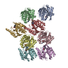
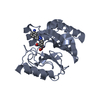
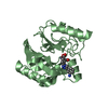
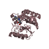
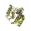
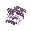
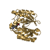
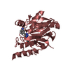
- PDB-7yhc: Crystal structure of VIM-2 MBL in complex with 3-(4-(3-aminopheny... -
+ Open data

- Basic information
| Entry | Database: PDB / ID: 7yhc | ||||||||||||
|---|---|---|---|---|---|---|---|---|---|---|---|---|---|
| Title | Crystal structure of VIM-2 MBL in complex with 3-(4-(3-aminophenyl)-1H-1,2,3-triazol-1-yl)phthalic acid | ||||||||||||
 Components Components | Beta-lactamase class B VIM-2 | ||||||||||||
 Keywords Keywords | HYDROLASE/INHIBITOR / Metallo-beta-lactamase VIM-2 / HYDROLASE / HYDROLASE-INHIBITOR complex | ||||||||||||
| Function / homology |  Function and homology information Function and homology informationantibiotic catabolic process / beta-lactamase activity / beta-lactamase / periplasmic space / response to antibiotic / metal ion binding Similarity search - Function | ||||||||||||
| Biological species |   Pseudomonas aeruginosa (bacteria) Pseudomonas aeruginosa (bacteria) | ||||||||||||
| Method |  X-RAY DIFFRACTION / SYNCHROTRON / MOLECULAR REPLACEMENT / molecular replacement / Resolution: 2.153 Å X-RAY DIFFRACTION / SYNCHROTRON / MOLECULAR REPLACEMENT / molecular replacement / Resolution: 2.153 Å | ||||||||||||
 Authors Authors | Li, G.-B. / Yan, Y.-H. | ||||||||||||
| Funding support |  China, 3items China, 3items
| ||||||||||||
 Citation Citation | Journal: Eur.J.Med.Chem. / Year: 2023 Title: Metal binding pharmacophore click-derived discovery of new broad-spectrum metallo-beta-lactamase inhibitors. Authors: Yan, Y.H. / Ding, H.S. / Zhu, K.R. / Mu, B.S. / Zheng, Y. / Huang, M.Y. / Zhou, C. / Li, W.F. / Wang, Z. / Wu, Y. / Li, G.B. | ||||||||||||
| History |
|
- Structure visualization
| Structure viewer | Molecule: MolmilJmol/JSmol |
|---|
- Downloads & links
-Download
| PDBx/mmCIF format | 7yhc.cif.gz | 365.8 KB | Display | PDBx/mmCIF format |
|---|---|---|---|---|
| PDB format | pdb7yhc.ent.gz | 297.9 KB | Display | PDB format |
| PDBx/mmJSON format | 7yhc.json.gz | Tree view | PDBx/mmJSON format | |
| Others |  Other downloads Other downloads |
-Validation report
| Arichive directory | https://data.pdbj.org/pub/pdb/validation_reports/yh/7yhcftp://data.pdbj.org/pub/pdb/validation_reports/yh/7yhc | HTTPS FTP |
|---|
-Related structure data
| Related structure data | 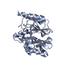 7yh9C  7yhaC 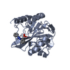 7yhbC 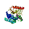 7yhdC  6jn6S S: Starting model for refinement C: citing same article ( |
|---|---|
| Similar structure data |
-Links
 PDBj
PDBj

- Assembly
-Components
| #1: Protein | Mass: 24679.439 Da / Num. of mol.: 8 Source method: isolated from a genetically manipulated source Details: Pseudomonas aeruginosa / Source: (gene. exp.) Pseudomonas aeruginosa (bacteria)Gene: blaVIM-2, bla vim-2, bla-VIM-2, blasVIM-2, blaVIM2, blm, VIM-2, vim-2, PAERUG_P32_London_17_VIM_2_10_11_06255 Production host: #2: Chemical | ChemComp-ZN /   Mass: 65.409 Da / Num. of mol.: 16 / Source method: obtained synthetically / Formula: Zn Mass: 65.409 Da / Num. of mol.: 16 / Source method: obtained synthetically / Formula: Zn#3: Chemical | ChemComp-IU3 / 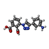  Mass: 324.291 Da / Num. of mol.: 8 / Source method: obtained synthetically / Formula: C16H12N4O4 / Feature type: SUBJECT OF INVESTIGATION Mass: 324.291 Da / Num. of mol.: 8 / Source method: obtained synthetically / Formula: C16H12N4O4 / Feature type: SUBJECT OF INVESTIGATION#4: Water | ChemComp-HOH / |  Mass: 18.015 Da / Num. of mol.: 653 / Source method: isolated from a natural source / Formula: H2O Mass: 18.015 Da / Num. of mol.: 653 / Source method: isolated from a natural source / Formula: H2OHas ligand of interest | Y | |
|---|
-Experimental details
-Experiment
| Experiment | Method: X-RAY DIFFRACTION / Number of used crystals: 1 |
|---|
- Sample preparation
| Crystal | Density Matthews: 2.21 Å3/Da / Density % sol: 44.35 % |
|---|---|
| Crystal grow | Temperature: 293 K / Method: vapor diffusion, hanging drop Details: 0.2 M Magnesium Formate, 23-30% (v/v) Polyethylene glycol 3350 |
-Data collection
| Diffraction | Mean temperature: 195 K / Serial crystal experiment: N |
|---|---|
| Diffraction source | Source: SYNCHROTRON / Site: SSRF / Beamline: BL19U1 / Wavelength: 1 Å |
| Detector | Type: DECTRIS PILATUS3 X CdTe 1M / Detector: PIXEL / Date: May 25, 2020 |
| Radiation | Protocol: SINGLE WAVELENGTH / Monochromatic (M) / Laue (L): M / Scattering type: x-ray |
| Radiation wavelength | Wavelength: 1 Å / Relative weight: 1 |
| Reflection | Resolution: 2.15→19.87 Å / Num. obs: 91180 / % possible obs: 98.95 % / Redundancy: 6.6 % / Biso Wilson estimate: 33.52 Å2 / CC1/2: 0.998 / Rmerge(I) obs: 0.074 / Net I/σ(I): 14.5 |
| Reflection shell | Resolution: 2.15→2.23 Å / Rmerge(I) obs: 0.346 / Num. unique obs: 8308 / CC1/2: 0.9 |
-Phasing
| Phasing | Method: molecular replacement | |||||||||
|---|---|---|---|---|---|---|---|---|---|---|
| Phasing MR |
|
- Processing
| Software |
| ||||||||||||||||||||||||||||||||||||||||||||||||||||||||||||||||||||||||||||||||||||||||||
|---|---|---|---|---|---|---|---|---|---|---|---|---|---|---|---|---|---|---|---|---|---|---|---|---|---|---|---|---|---|---|---|---|---|---|---|---|---|---|---|---|---|---|---|---|---|---|---|---|---|---|---|---|---|---|---|---|---|---|---|---|---|---|---|---|---|---|---|---|---|---|---|---|---|---|---|---|---|---|---|---|---|---|---|---|---|---|---|---|---|---|---|
| Refinement | Method to determine structure: MOLECULAR REPLACEMENT Starting model: 6JN6 Resolution: 2.153→19.828 Å / SU ML: 0.25 / Cross valid method: THROUGHOUT / σ(F): 1.74 / Phase error: 23.23 / Stereochemistry target values: ML
| ||||||||||||||||||||||||||||||||||||||||||||||||||||||||||||||||||||||||||||||||||||||||||
| Solvent computation | Shrinkage radii: 0.9 Å / VDW probe radii: 1.11 Å / Solvent model: FLAT BULK SOLVENT MODEL | ||||||||||||||||||||||||||||||||||||||||||||||||||||||||||||||||||||||||||||||||||||||||||
| Displacement parameters | Biso max: 81.76 Å2 / Biso mean: 35.6167 Å2 / Biso min: 17.08 Å2 | ||||||||||||||||||||||||||||||||||||||||||||||||||||||||||||||||||||||||||||||||||||||||||
| Refinement step | Cycle: final / Resolution: 2.153→19.828 Å
| ||||||||||||||||||||||||||||||||||||||||||||||||||||||||||||||||||||||||||||||||||||||||||
| Refine LS restraints |
| ||||||||||||||||||||||||||||||||||||||||||||||||||||||||||||||||||||||||||||||||||||||||||
| LS refinement shell | Refine-ID: X-RAY DIFFRACTION / Rfactor Rfree error: 0
|
