ripoptosome assembly / positive regulation of miRNA processing / positive regulation of interleukin-6-mediated signaling pathway / death domain binding / ripoptosome assembly involved in necroptotic process / peptidyl-serine autophosphorylation / Defective RIPK1-mediated regulated necrosis / Microbial modulation of RIPK1-mediated regulated necrosis / ripoptosome / TRIF-mediated programmed cell death ...ripoptosome assembly / positive regulation of miRNA processing / positive regulation of interleukin-6-mediated signaling pathway / death domain binding / ripoptosome assembly involved in necroptotic process / peptidyl-serine autophosphorylation / Defective RIPK1-mediated regulated necrosis / Microbial modulation of RIPK1-mediated regulated necrosis / ripoptosome / TRIF-mediated programmed cell death / Regulation by c-FLIP / CASP8 activity is inhibited / Dimerization of procaspase-8 / TLR3-mediated TICAM1-dependent programmed cell death / programmed necrotic cell death / TNF signaling / SARS-CoV-1-mediated effects on programmed cell death / Caspase activation via Death Receptors in the presence of ligand / T cell apoptotic process / positive regulation of macrophage differentiation / JUN kinase kinase kinase activity / necroptotic signaling pathway / NF-kB activation through FADD/RIP-1 pathway mediated by caspase-8 and -10 / RIP-mediated NFkB activation via ZBP1 / death-inducing signaling complex / positive regulation of necroptotic process / negative regulation of necroptotic process / positive regulation of tumor necrosis factor-mediated signaling pathway / death receptor binding / positive regulation of programmed cell death / positive regulation of programmed necrotic cell death / TNFR1-induced proapoptotic signaling / positive regulation of extrinsic apoptotic signaling pathway / RIPK1-mediated regulated necrosis / TRP channels / necroptotic process / response to tumor necrosis factor / negative regulation of extrinsic apoptotic signaling pathway in absence of ligand / positive regulation of execution phase of apoptosis / extrinsic apoptotic signaling pathway / canonical NF-kappaB signal transduction / signaling adaptor activity / TICAM1, RIP1-mediated IKK complex recruitment / tumor necrosis factor-mediated signaling pathway / : / negative regulation of extrinsic apoptotic signaling pathway / positive regulation of interleukin-8 production / protein serine/threonine kinase binding / IKK complex recruitment mediated by RIP1 / protein catabolic process / TNFR1-induced NF-kappa-B signaling pathway / negative regulation of canonical NF-kappaB signal transduction / Regulation of TNFR1 signaling / positive regulation of non-canonical NF-kappaB signal transduction / positive regulation of protein phosphorylation / positive regulation of JNK cascade / Regulation of necroptotic cell death / cellular response to growth factor stimulus / cellular response to tumor necrosis factor / positive regulation of reactive oxygen species metabolic process / cellular response to hydrogen peroxide / positive regulation of tumor necrosis factor production / positive regulation of inflammatory response / Ovarian tumor domain proteases / protein autophosphorylation / positive regulation of neuron apoptotic process / response to oxidative stress / amyloid fibril formation / Potential therapeutics for SARS / protein kinase activity / positive regulation of canonical NF-kappaB signal transduction / non-specific serine/threonine protein kinase / signaling receptor complex / endosome membrane / Ub-specific processing proteases / intracellular signal transduction / positive regulation of apoptotic process / inflammatory response / protein serine kinase activity / protein serine/threonine kinase activity / apoptotic process / ubiquitin protein ligase binding / positive regulation of gene expression / negative regulation of apoptotic process / protein-containing complex binding / protein homodimerization activity / positive regulation of transcription by RNA polymerase II / protein-containing complex / mitochondrion / ATP binding / identical protein binding / plasma membrane / cytoplasm / cytosol Similarity search - Function
RIP1, Death domain / Death domain profile. / DEATH domain, found in proteins involved in cell death (apoptosis). / Death domain / Death domain / : / Death-like domain superfamily / Protein tyrosine and serine/threonine kinase / Serine-threonine/tyrosine-protein kinase, catalytic domain / Serine/threonine-protein kinase, active site ...RIP1, Death domain / Death domain profile. / DEATH domain, found in proteins involved in cell death (apoptosis). / Death domain / Death domain / : / Death-like domain superfamily / Protein tyrosine and serine/threonine kinase / Serine-threonine/tyrosine-protein kinase, catalytic domain / Serine/threonine-protein kinase, active site / Serine/Threonine protein kinases active-site signature. / Serine/Threonine protein kinases, catalytic domain / Protein kinase domain profile. / Protein kinase domain / Protein kinase-like domain superfamily Similarity search - Domain/homology
In the structure databanks used in Yorodumi, some data are registered as the other names, "COVID-19 virus" and "2019-nCoV". Here are the details of the virus and the list of structure data.
Jan 31, 2019. EMDB accession codes are about to change! (news from PDBe EMDB page)
EMDB accession codes are about to change! (news from PDBe EMDB page)
The allocation of 4 digits for EMDB accession codes will soon come to an end. Whilst these codes will remain in use, new EMDB accession codes will include an additional digit and will expand incrementally as the available range of codes is exhausted. The current 4-digit format prefixed with “EMD-” (i.e. EMD-XXXX) will advance to a 5-digit format (i.e. EMD-XXXXX), and so on. It is currently estimated that the 4-digit codes will be depleted around Spring 2019, at which point the 5-digit format will come into force.
The EM Navigator/Yorodumi systems omit the EMD- prefix.
Related info.:Q: What is EMD? / ID/Accession-code notation in Yorodumi/EM Navigator
Yorodumi is a browser for structure data from EMDB, PDB, SASBDB, etc.
This page is also the successor to EM Navigator detail page, and also detail information page/front-end page for Omokage search.
The word "yorodu" (or yorozu) is an old Japanese word meaning "ten thousand". "mi" (miru) is to see.
Related info.:EMDB / PDB / SASBDB / Comparison of 3 databanks / Yorodumi Search / Aug 31, 2016. New EM Navigator & Yorodumi / Yorodumi Papers / Jmol/JSmol / Function and homology information / Changes in new EM Navigator and Yorodumi
 Movie
Movie Controller

 Yorodumi
 Open data
 Basic information
 Components
 Keywords
 Function and homology information
 Homo sapiens (human)
 X-RAY DIFFRACTION /
  Authors
 Citation
 Structure visualization
 Downloads & links
 Other downloads
 PDBj
PDBj


 Assembly
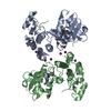
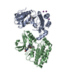

 Spodoptera frugiperda (fall armyworm)
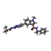
 Mass: 480.561 Da / Num. of mol.: 2 / Source method: obtained synthetically / Formula: C28H28N6O2 / Feature type: SUBJECT OF INVESTIGATION
 Mass: 126.904 Da / Num. of mol.: 7 / Source method: obtained synthetically / Formula: I
 Mass: 18.015 Da / Num. of mol.: 65 / Source method: isolated from a natural source / Formula: H2O
 Sample preparation
 / Beamline: BL19U1 / Wavelength: 0.97852 Å
 Processing