 Movie
Movie Controller

[English] 日本語
 Yorodumi
- PDB-7ych: Cryo-EM structure of Tetrahymena ribozyme conformation 3 undergoi... -
+ Open data

- Basic information
| Entry | Database: PDB / ID: 7ych | ||||||
|---|---|---|---|---|---|---|---|
| Title | Cryo-EM structure of Tetrahymena ribozyme conformation 3 undergoing the first-step self-splicing | ||||||
 Components Components |
| ||||||
 Keywords Keywords | RNA / Tetrahymena ribozyme / first step of self-splicing / conformation 3 | ||||||
| Function / homology | GUANOSINE-5'-TRIPHOSPHATE / : / RNA / RNA (> 10) / RNA (> 100) Function and homology information Function and homology information | ||||||
| Biological species |  Tetrahymena (eukaryote) Tetrahymena (eukaryote) | ||||||
| Method | ELECTRON MICROSCOPY / single particle reconstruction / cryo EM / Resolution: 3.09 Å | ||||||
 Authors Authors | Zhang, X. / Li, S. / Pintilie, G. / Palo, M.Z. / Zhang, K. | ||||||
| Funding support |  China, 1items China, 1items
| ||||||
 Citation Citation | Journal: Nucleic Acids Res / Year: 2023 Title: Snapshots of the first-step self-splicing of Tetrahymena ribozyme revealed by cryo-EM. Authors: Xiaojing Zhang / Shanshan Li / Grigore Pintilie / Michael Z Palo / Kaiming Zhang /  Abstract: Tetrahymena ribozyme is a group I intron, whose self-splicing is the result of two sequential ester-transfer reactions. To understand how it facilitates catalysis in the first self-splicing reaction, ...Tetrahymena ribozyme is a group I intron, whose self-splicing is the result of two sequential ester-transfer reactions. To understand how it facilitates catalysis in the first self-splicing reaction, we used cryogenic electron microscopy (cryo-EM) to resolve the structures of L-16 Tetrahymena ribozyme complexed with a 11-nucleotide 5'-splice site analog substrate. Four conformations were achieved to 4.14, 3.18, 3.09 and 2.98 Å resolutions, respectively, corresponding to different splicing intermediates during the first enzymatic reaction. Comparison of these structures reveals structural alterations, including large conformational changes in IGS/IGSext (P1-P1ext duplex) and J5/4, as well as subtle local rearrangements in the G-binding site. These structural changes are required for the enzymatic activity of the Tetrahymena ribozyme. Our study demonstrates the ability of cryo-EM to capture dynamic RNA structural changes, ushering in a new era in the analysis of RNA structure-function by cryo-EM. | ||||||
| History |
|
- Structure visualization
| Structure viewer | Molecule: MolmilJmol/JSmol |
|---|
- Downloads & links
-Download
| PDBx/mmCIF format | 7ych.cif.gz | 203.9 KB | Display | PDBx/mmCIF format |
|---|---|---|---|---|
| PDB format | pdb7ych.ent.gz | 148.9 KB | Display | PDB format |
| PDBx/mmJSON format | 7ych.json.gz | Tree view | PDBx/mmJSON format | |
| Others |  Other downloads Other downloads |
-Validation report
| Summary document | 7ych_validation.pdf.gz | 1.3 MB | Display | wwPDB validaton report |
|---|---|---|---|---|
| Full document | 7ych_full_validation.pdf.gz | 1.4 MB | Display | |
| Data in XML | 7ych_validation.xml.gz | 23.4 KB | Display | |
| Data in CIF | 7ych_validation.cif.gz | 33.2 KB | Display | |
| Arichive directory | https://data.pdbj.org/pub/pdb/validation_reports/yc/7ychftp://data.pdbj.org/pub/pdb/validation_reports/yc/7ych | HTTPS FTP |
-Related structure data
| Related structure data |  33739MC  7yc8C 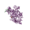 7ycgC 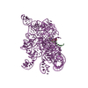 7yciC M: map data used to model this data C: citing same article ( |
|---|---|
| Similar structure data |


-Links
 PDBj
PDBj


- Assembly
| Deposited unit | 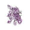
|
|---|---|
| 1 |
|
-Components
| #1: RNA chain | Mass: 3386.082 Da / Num. of mol.: 1 / Source method: obtained synthetically / Source: (synth.) Tetrahymena (eukaryote) | ||
|---|---|---|---|
| #2: RNA chain | Mass: 127011.883 Da / Num. of mol.: 1 Source method: isolated from a genetically manipulated source Source: (gene. exp.) Tetrahymena (eukaryote)Production host: in vitro transcription vector pT7-Fluc(deltai) (others) References: GenBank: 10832 | ||
| #3: Chemical | ChemComp-GTP /   Mass: 523.180 Da / Num. of mol.: 1 / Source method: obtained synthetically / Formula: C10H16N5O14P3 / Feature type: SUBJECT OF INVESTIGATION / Comment: GTP, energy-carrying molecule*YM Mass: 523.180 Da / Num. of mol.: 1 / Source method: obtained synthetically / Formula: C10H16N5O14P3 / Feature type: SUBJECT OF INVESTIGATION / Comment: GTP, energy-carrying molecule*YM | ||
| #4: Chemical |   Mass: 24.305 Da / Num. of mol.: 3 / Source method: obtained synthetically / Formula: Mg / Feature type: SUBJECT OF INVESTIGATION Mass: 24.305 Da / Num. of mol.: 3 / Source method: obtained synthetically / Formula: Mg / Feature type: SUBJECT OF INVESTIGATIONHas ligand of interest | Y | |
-Experimental details
-Experiment
| Experiment | Method: ELECTRON MICROSCOPY |
|---|---|
| EM experiment | Aggregation state: PARTICLE / 3D reconstruction method: single particle reconstruction |
- Sample preparation
| Component | Name: Cryo-EM structure of Tetrahymena ribozyme conformation 3 undergoing the first-step self-splicing Type: COMPLEX / Entity ID: #1-#2 / Source: RECOMBINANT |
|---|---|
| Molecular weight | Value: 0.13 MDa / Experimental value: YES |
| Source (natural) | Organism: Tetrahymena (eukaryote) |
| Source (recombinant) | Organism: in vitro transcription vector pT7-Fluc(deltai) (others) |
| Buffer solution | pH: 7.5 |
| Specimen | Conc.: 1 mg/ml / Embedding applied: NO / Shadowing applied: NO / Staining applied: NO / Vitrification applied: YES |
| Vitrification | Cryogen name: ETHANE |
- Electron microscopy imaging
| Experimental equipment |  Model: Titan Krios / Image courtesy: FEI Company |
|---|---|
| Microscopy | Model: FEI TITAN KRIOS |
| Electron gun | Electron source:  FIELD EMISSION GUN / Accelerating voltage: 300 kV / Illumination mode: FLOOD BEAM FIELD EMISSION GUN / Accelerating voltage: 300 kV / Illumination mode: FLOOD BEAM |
| Electron lens | Mode: BRIGHT FIELD / Nominal defocus max: 2800 nm / Nominal defocus min: 800 nm |
| Image recording | Electron dose: 51 e/Å2 / Film or detector model: GATAN K3 (6k x 4k) |
- Processing
| EM software |
| ||||||||||||||||
|---|---|---|---|---|---|---|---|---|---|---|---|---|---|---|---|---|---|
| CTF correction | Type: NONE | ||||||||||||||||
| Particle selection | Num. of particles selected: 3668247 | ||||||||||||||||
| 3D reconstruction | Resolution: 3.09 Å / Resolution method: FSC 0.143 CUT-OFF / Num. of particles: 202401 / Symmetry type: POINT |
