peptide-aspartate beta-dioxygenase / : / regulation of protein depolymerization / activation of store-operated calcium channel activity / regulation of cell communication by electrical coupling / junctional sarcoplasmic reticulum membrane / peptidyl-aspartic acid 3-dioxygenase activity / sarcoplasmic reticulum lumen / cortical endoplasmic reticulum / positive regulation of intracellular protein transport ...peptide-aspartate beta-dioxygenase / : / regulation of protein depolymerization / activation of store-operated calcium channel activity / regulation of cell communication by electrical coupling / junctional sarcoplasmic reticulum membrane / peptidyl-aspartic acid 3-dioxygenase activity / sarcoplasmic reticulum lumen / cortical endoplasmic reticulum / positive regulation of intracellular protein transport / limb morphogenesis / pattern specification process / face morphogenesis / structural constituent of muscle / response to ATP / positive regulation of proteolysis / roof of mouth development / Protein hydroxylation / positive regulation of calcium ion transport into cytosol / detection of calcium ion / regulation of cytosolic calcium ion concentration / calcium ion homeostasis / regulation of release of sequestered calcium ion into cytosol by sarcoplasmic reticulum / Ion homeostasis / regulation of cardiac muscle contraction by regulation of the release of sequestered calcium ion / sarcoplasmic reticulum membrane / calcium channel complex / cellular response to calcium ion / muscle contraction / regulation of protein stability / Stimuli-sensing channels / calcium ion transmembrane transport / transmembrane transporter binding / electron transfer activity / cell population proliferation / negative regulation of cell population proliferation / calcium ion binding / endoplasmic reticulum membrane / positive regulation of DNA-templated transcription / structural molecule activity / endoplasmic reticulum / plasma membrane Similarity search - Function
In the structure databanks used in Yorodumi, some data are registered as the other names, "COVID-19 virus" and "2019-nCoV". Here are the details of the virus and the list of structure data.
Jan 31, 2019. EMDB accession codes are about to change! (news from PDBe EMDB page)
EMDB accession codes are about to change! (news from PDBe EMDB page)
The allocation of 4 digits for EMDB accession codes will soon come to an end. Whilst these codes will remain in use, new EMDB accession codes will include an additional digit and will expand incrementally as the available range of codes is exhausted. The current 4-digit format prefixed with “EMD-” (i.e. EMD-XXXX) will advance to a 5-digit format (i.e. EMD-XXXXX), and so on. It is currently estimated that the 4-digit codes will be depleted around Spring 2019, at which point the 5-digit format will come into force.
The EM Navigator/Yorodumi systems omit the EMD- prefix.
Related info.:Q: What is EMD? / ID/Accession-code notation in Yorodumi/EM Navigator
Yorodumi is a browser for structure data from EMDB, PDB, SASBDB, etc.
This page is also the successor to EM Navigator detail page, and also detail information page/front-end page for Omokage search.
The word "yorodu" (or yorozu) is an old Japanese word meaning "ten thousand". "mi" (miru) is to see.
Related info.:EMDB / PDB / SASBDB / Comparison of 3 databanks / Yorodumi Search / Aug 31, 2016. New EM Navigator & Yorodumi / Yorodumi Papers / Jmol/JSmol / Function and homology information / Changes in new EM Navigator and Yorodumi
 Movie
Movie Controller

 Yorodumi
 Open data
 Basic information
 Components
 Keywords
 Function and homology information
 Homo sapiens (human)
 X-RAY DIFFRACTION /
  Authors
 United Kingdom, 1items
  Citation
 Structure visualization
 Downloads & links
 Other downloads
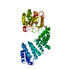
 PDBj
PDBj


 Assembly
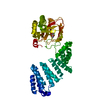


 Mass: 54.938 Da / Num. of mol.: 1 / Source method: obtained synthetically / Formula: Mn
 Mass: 162.098 Da / Num. of mol.: 1 / Source method: obtained synthetically / Formula: C5H6O6 / Feature type: SUBJECT OF INVESTIGATION
 Mass: 18.015 Da / Num. of mol.: 222 / Source method: isolated from a natural source / Formula: H2O
 Sample preparation
 Processing