 Movie
Movie Controller

[English] 日本語
 Yorodumi
- PDB-7ya2: Crystal structure of capsular polysaccharide synthesis enzyme Cap... -
+ Open data

- Basic information
| Entry | Database: PDB / ID: 7ya2 | ||||||
|---|---|---|---|---|---|---|---|
| Title | Crystal structure of capsular polysaccharide synthesis enzyme CapG from Staphylococcus aureus | ||||||
 Components Components | Capsular polysaccharide synthesis enzyme CapG | ||||||
 Keywords Keywords | ISOMERASE / 2-epimerase | ||||||
| Function / homology | UDP-N-acetylglucosamine 2-epimerase WecB-like / UDP-N-acetylglucosamine 2-epimerase activity / UDP-N-acetylglucosamine 2-epimerase domain / UDP-N-acetylglucosamine 2-epimerase / Capsular polysaccharide synthesis enzyme CapG Function and homology information Function and homology information | ||||||
| Biological species |   Staphylococcus aureus (bacteria) Staphylococcus aureus (bacteria) | ||||||
| Method |  X-RAY DIFFRACTION / SYNCHROTRON / MOLECULAR REPLACEMENT / Resolution: 3.2 Å X-RAY DIFFRACTION / SYNCHROTRON / MOLECULAR REPLACEMENT / Resolution: 3.2 Å | ||||||
 Authors Authors | Chen, Y. / Wang, Y.C. | ||||||
| Funding support |  Taiwan, 1items Taiwan, 1items
| ||||||
 Citation Citation | Journal: Acta Crystallogr.,Sect.F / Year: 2022 Title: Crystal structure of the capsular polysaccharide-synthesis enzyme CapG from Staphylococcus aureus. Authors: Tien, N. / Ho, C.Y. / Lai, S.J. / Lin, Y.C. / Yang, C.S. / Wang, Y.C. / Huang, W.C. / Chen, Y. / Chang, J.J. | ||||||
| History |
|
- Structure visualization
| Structure viewer | Molecule: MolmilJmol/JSmol |
|---|
- Downloads & links
-Download
| PDBx/mmCIF format | 7ya2.cif.gz | 444.3 KB | Display | PDBx/mmCIF format |
|---|---|---|---|---|
| PDB format | pdb7ya2.ent.gz | 363.7 KB | Display | PDB format |
| PDBx/mmJSON format | 7ya2.json.gz | Tree view | PDBx/mmJSON format | |
| Others |  Other downloads Other downloads |
-Validation report
| Arichive directory | https://data.pdbj.org/pub/pdb/validation_reports/ya/7ya2ftp://data.pdbj.org/pub/pdb/validation_reports/ya/7ya2 | HTTPS FTP |
|---|
-Related structure data
| Related structure data | 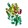 4hwgS S: Starting model for refinement |
|---|---|
| Similar structure data |
-Links
 PDBj
PDBj- Assembly
| Deposited unit | 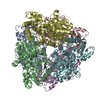
| ||||||||||||
|---|---|---|---|---|---|---|---|---|---|---|---|---|---|
| 1 |
| ||||||||||||
| Unit cell |
|
-Components
| #1: Protein | Mass: 42905.785 Da / Num. of mol.: 6 Source method: isolated from a genetically manipulated source Source: (gene. exp.) Staphylococcus aureus (bacteria) / Strain: Newman / Gene: capG, NWMN_0101, CNH35_00585 / Production host: |
|---|
-Experimental details
-Experiment
| Experiment | Method: X-RAY DIFFRACTION / Number of used crystals: 1 |
|---|
- Sample preparation
| Crystal | Density Matthews: 3.37 Å3/Da / Density % sol: 63.48 % |
|---|---|
| Crystal grow | Temperature: 277 K / Method: vapor diffusion, sitting drop Details: 0.2 M ammonium citrate tribasic pH 7.2, 18 % w/v polyethylene glycol 3350 |
-Data collection
| Diffraction | Mean temperature: 100 K / Serial crystal experiment: N |
|---|---|
| Diffraction source | Source: SYNCHROTRON / Site: NSRRC / Beamline: BL13B1 / Wavelength: 1 Å |
| Detector | Type: ADSC QUANTUM 315r / Detector: CCD / Date: Apr 13, 2022 |
| Radiation | Protocol: SINGLE WAVELENGTH / Monochromatic (M) / Laue (L): M / Scattering type: x-ray |
| Radiation wavelength | Wavelength: 1 Å / Relative weight: 1 |
| Reflection | Resolution: 3.2→30 Å / Num. obs: 56178 / % possible obs: 99.4 % / Redundancy: 3.5 % / Rmerge(I) obs: 0.126 / Net I/σ(I): 9.675 |
| Reflection shell | Resolution: 3.2→3.31 Å / Redundancy: 3.4 % / Num. unique obs: 5591 / CC1/2: 0.591 / CC star: 0.862 / % possible all: 99.8 |
- Processing
| Software |
| ||||||||||||||||||||||||
|---|---|---|---|---|---|---|---|---|---|---|---|---|---|---|---|---|---|---|---|---|---|---|---|---|---|
| Refinement | Method to determine structure: MOLECULAR REPLACEMENT Starting model: 4HWG Resolution: 3.2→25.57 Å / Cross valid method: THROUGHOUT Stereochemistry target values: GeoStd + Monomer Library + CDL v1.2
| ||||||||||||||||||||||||
| Displacement parameters | Biso mean: 74.66 Å2 | ||||||||||||||||||||||||
| Refinement step | Cycle: LAST / Resolution: 3.2→25.57 Å
| ||||||||||||||||||||||||
| Refine LS restraints |
|
