 Movie
Movie Controller


+ Open data

- Basic information
| Entry | Database: PDB / ID: 7y8a | ||||||
|---|---|---|---|---|---|---|---|
| Title | Cryo-EM structure of cryptophyte photosystem I | ||||||
 Components Components |
| ||||||
 Keywords Keywords | PHOTOSYNTHESIS / Cryptophyte / Photosystem I / evolution | ||||||
| Function / homology |  Function and homology information Function and homology informationplastid thylakoid membrane / thylakoid membrane / photosystem I reaction center / photosystem I / photosynthetic electron transport in photosystem I / photosystem I / chlorophyll binding / chloroplast thylakoid membrane / photosynthesis / 4 iron, 4 sulfur cluster binding ...plastid thylakoid membrane / thylakoid membrane / photosystem I reaction center / photosystem I / photosynthetic electron transport in photosystem I / photosystem I / chlorophyll binding / chloroplast thylakoid membrane / photosynthesis / 4 iron, 4 sulfur cluster binding / electron transfer activity / oxidoreductase activity / magnesium ion binding / metal ion binding Similarity search - Function | ||||||
| Biological species |  Chroomonas placoidea (eukaryote) Chroomonas placoidea (eukaryote) | ||||||
| Method | ELECTRON MICROSCOPY / single particle reconstruction / cryo EM / Resolution: 2.71 Å | ||||||
 Authors Authors | Zhao, L.S. / Zhang, Y.Z. / Liu, L.N. / Li, K. | ||||||
| Funding support |  China, 1items China, 1items
| ||||||
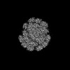
 Citation Citation | Journal: Plant Cell / Year: 2023 Title: Structural basis and evolution of the photosystem I-light-harvesting supercomplex of cryptophyte algae. Authors: Long-Sheng Zhao / Peng Wang / Kang Li / Quan-Bao Zhang / Fei-Yu He / Chun-Yang Li / Hai-Nan Su / Xiu-Lan Chen / Lu-Ning Liu / Yu-Zhong Zhang /  Abstract: Cryptophyte plastids originated from a red algal ancestor through secondary endosymbiosis. Cryptophyte photosystem I (PSI) associates with transmembrane alloxanthin-chlorophyll a/c proteins (ACPIs) ...Cryptophyte plastids originated from a red algal ancestor through secondary endosymbiosis. Cryptophyte photosystem I (PSI) associates with transmembrane alloxanthin-chlorophyll a/c proteins (ACPIs) as light-harvesting complexes (LHCs). Here, we report the structure of the photosynthetic PSI-ACPI supercomplex from the cryptophyte Chroomonas placoidea at 2.7-Å resolution obtained by crygenic electron microscopy. Cryptophyte PSI-ACPI represents a unique PSI-LHCI intermediate in the evolution from red algal to diatom PSI-LHCI. The PSI-ACPI supercomplex is composed of a monomeric PSI core containing 14 subunits, 12 of which originated in red algae, 1 diatom PsaR homolog, and an additional peptide. The PSI core is surrounded by 14 ACPI subunits that form 2 antenna layers: an inner layer with 11 ACPIs surrounding the PSI core and an outer layer containing 3 ACPIs. A pigment-binding subunit that is not present in any other previously characterized PSI-LHCI complexes, ACPI-S, mediates the association and energy transfer between the outer and inner ACPIs. The extensive pigment network of PSI-ACPI ensures efficient light harvesting, energy transfer, and dissipation. Overall, the PSI-LHCI structure identified in this study provides a framework for delineating the mechanisms of energy transfer in cryptophyte PSI-LHCI and for understanding the evolution of photosynthesis in the red lineage, which occurred via secondary endosymbiosis. | ||||||
| History |
|
- Structure visualization
| Structure viewer | Molecule: MolmilJmol/JSmol |
|---|
- Downloads & links
-Download
| PDBx/mmCIF format | 7y8a.cif.gz | 1.3 MB | Display | PDBx/mmCIF format |
|---|---|---|---|---|
| PDB format | pdb7y8a.ent.gz | 1.2 MB | Display | PDB format |
| PDBx/mmJSON format | 7y8a.json.gz | Tree view | PDBx/mmJSON format | |
| Others |  Other downloads Other downloads |
-Validation report
| Arichive directory | https://data.pdbj.org/pub/pdb/validation_reports/y8/7y8aftp://data.pdbj.org/pub/pdb/validation_reports/y8/7y8a | HTTPS FTP |
|---|
-Related structure data
| Related structure data | 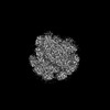 33683MC 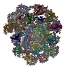 7y7bC M: map data used to model this data C: citing same article ( |
|---|---|
| Similar structure data |
-Links
 PDBj
PDBj


- Assembly
| Deposited unit | 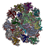
|
|---|---|
| 1 |
|
-Components
-Protein , 16 types, 16 molecules 123456789CORXZab
| #1: Protein | Mass: 23479.367 Da / Num. of mol.: 1 / Source method: isolated from a natural source / Source: (natural) Chroomonas placoidea (eukaryote) |
|---|---|
| #2: Protein | Mass: 23400.123 Da / Num. of mol.: 1 / Source method: isolated from a natural source / Source: (natural) Chroomonas placoidea (eukaryote) |
| #3: Protein | Mass: 24966.262 Da / Num. of mol.: 1 / Source method: isolated from a natural source / Source: (natural) Chroomonas placoidea (eukaryote) |
| #4: Protein | Mass: 23548.246 Da / Num. of mol.: 1 / Source method: isolated from a natural source / Source: (natural) Chroomonas placoidea (eukaryote) |
| #5: Protein | Mass: 24055.031 Da / Num. of mol.: 1 / Source method: isolated from a natural source / Source: (natural) Chroomonas placoidea (eukaryote) |
| #6: Protein | Mass: 22688.564 Da / Num. of mol.: 1 / Source method: isolated from a natural source / Source: (natural) Chroomonas placoidea (eukaryote) |
| #7: Protein | Mass: 24550.402 Da / Num. of mol.: 1 / Source method: isolated from a natural source / Source: (natural) Chroomonas placoidea (eukaryote) |
| #8: Protein | Mass: 23700.525 Da / Num. of mol.: 1 / Source method: isolated from a natural source / Source: (natural) Chroomonas placoidea (eukaryote) |
| #9: Protein | Mass: 23223.098 Da / Num. of mol.: 1 / Source method: isolated from a natural source / Source: (natural) Chroomonas placoidea (eukaryote) |
| #12: Protein | Mass: 8743.131 Da / Num. of mol.: 1 / Source method: isolated from a natural source / Source: (natural) Chroomonas placoidea (eukaryote) / References: UniProt: A0A222AI25, photosystem I |
| #21: Protein | Mass: 16253.030 Da / Num. of mol.: 1 / Source method: isolated from a natural source / Source: (natural) Chroomonas placoidea (eukaryote) |
| #22: Protein | Mass: 13867.048 Da / Num. of mol.: 1 / Source method: isolated from a natural source / Source: (natural) Chroomonas placoidea (eukaryote) |
| #23: Protein | Mass: 13975.214 Da / Num. of mol.: 1 / Source method: isolated from a natural source Details: Authors do not know how the coordinates align with the sequence and the residue numbering is arbitrary. Source: (natural) Chroomonas placoidea (eukaryote) |
| #24: Protein | Mass: 25645.455 Da / Num. of mol.: 1 / Source method: isolated from a natural source / Source: (natural) Chroomonas placoidea (eukaryote) |
| #25: Protein | Mass: 22270.992 Da / Num. of mol.: 1 / Source method: isolated from a natural source / Source: (natural) Chroomonas placoidea (eukaryote) |
| #26: Protein | Mass: 23755.605 Da / Num. of mol.: 1 / Source method: isolated from a natural source / Source: (natural) Chroomonas placoidea (eukaryote) |
-Photosystem I P700 chlorophyll a apoprotein ... , 2 types, 2 molecules AB
| #10: Protein | Mass: 83493.570 Da / Num. of mol.: 1 / Source method: isolated from a natural source / Source: (natural) Chroomonas placoidea (eukaryote) / References: UniProt: A0A222AIB4, photosystem I |
|---|---|
| #11: Protein | Mass: 82219.820 Da / Num. of mol.: 1 / Source method: isolated from a natural source / Source: (natural) Chroomonas placoidea (eukaryote) / References: UniProt: A0A222AI95, photosystem I |
-Photosystem I reaction center subunit ... , 8 types, 8 molecules DEFIJKLM
| #13: Protein | Mass: 15590.765 Da / Num. of mol.: 1 / Source method: isolated from a natural source / Source: (natural) Chroomonas placoidea (eukaryote) / References: UniProt: A0A222AIA6 |
|---|---|
| #14: Protein | Mass: 7352.387 Da / Num. of mol.: 1 / Source method: isolated from a natural source / Source: (natural) Chroomonas placoidea (eukaryote) / References: UniProt: A0A222AIF3 |
| #15: Protein | Mass: 20393.484 Da / Num. of mol.: 1 / Source method: isolated from a natural source / Source: (natural) Chroomonas placoidea (eukaryote) / References: UniProt: A0A222AI52 |
| #16: Protein/peptide | Mass: 3975.751 Da / Num. of mol.: 1 / Source method: isolated from a natural source / Source: (natural) Chroomonas placoidea (eukaryote) / References: UniProt: A0A222AI78 |
| #17: Protein/peptide | Mass: 4862.727 Da / Num. of mol.: 1 / Source method: isolated from a natural source / Source: (natural) Chroomonas placoidea (eukaryote) / References: UniProt: A0A222AI55 |
| #18: Protein | Mass: 8836.371 Da / Num. of mol.: 1 / Source method: isolated from a natural source / Source: (natural) Chroomonas placoidea (eukaryote) / References: UniProt: A0A222AI34 |
| #19: Protein | Mass: 16490.883 Da / Num. of mol.: 1 / Source method: isolated from a natural source / Source: (natural) Chroomonas placoidea (eukaryote) / References: UniProt: A0A222AI68 |
| #20: Protein/peptide | Mass: 3247.952 Da / Num. of mol.: 1 / Source method: isolated from a natural source / Source: (natural) Chroomonas placoidea (eukaryote) / References: UniProt: A0A222AI28 |
-Sugars , 2 types, 5 molecules 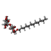

| #36: Sugar |  Type: D-saccharide / Mass: 510.615 Da / Num. of mol.: 3 / Source method: obtained synthetically / Formula: C24H46O11 / Comment: detergent*YM Type: D-saccharide / Mass: 510.615 Da / Num. of mol.: 3 / Source method: obtained synthetically / Formula: C24H46O11 / Comment: detergent*YM#39: Sugar |  Type: saccharide / Mass: 949.299 Da / Num. of mol.: 2 / Source method: obtained synthetically / Formula: C51H96O15 Type: saccharide / Mass: 949.299 Da / Num. of mol.: 2 / Source method: obtained synthetically / Formula: C51H96O15 |
|---|
-Non-polymers , 11 types, 352 molecules 


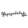


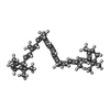


| #27: Chemical | ChemComp-CLA /  Mass: 893.489 Da / Num. of mol.: 222 / Source method: obtained synthetically / Formula: C55H72MgN4O5 / Feature type: SUBJECT OF INVESTIGATION Mass: 893.489 Da / Num. of mol.: 222 / Source method: obtained synthetically / Formula: C55H72MgN4O5 / Feature type: SUBJECT OF INVESTIGATION#28: Chemical | ChemComp-KC2 /  Mass: 608.926 Da / Num. of mol.: 14 / Source method: obtained synthetically / Formula: C35H28MgN4O5 / Feature type: SUBJECT OF INVESTIGATION Mass: 608.926 Da / Num. of mol.: 14 / Source method: obtained synthetically / Formula: C35H28MgN4O5 / Feature type: SUBJECT OF INVESTIGATION#29: Chemical | ChemComp-II0 / (  Mass: 564.840 Da / Num. of mol.: 47 / Source method: obtained synthetically / Formula: C40H52O2 / Feature type: SUBJECT OF INVESTIGATION Mass: 564.840 Da / Num. of mol.: 47 / Source method: obtained synthetically / Formula: C40H52O2 / Feature type: SUBJECT OF INVESTIGATION#30: Chemical |  Mass: 566.856 Da / Num. of mol.: 2 / Source method: obtained synthetically / Formula: C40H54O2 / Feature type: SUBJECT OF INVESTIGATION Mass: 566.856 Da / Num. of mol.: 2 / Source method: obtained synthetically / Formula: C40H54O2 / Feature type: SUBJECT OF INVESTIGATION#31: Chemical | ChemComp-IHT / (  Mass: 550.856 Da / Num. of mol.: 10 / Source method: obtained synthetically / Formula: C40H54O / Feature type: SUBJECT OF INVESTIGATION Mass: 550.856 Da / Num. of mol.: 10 / Source method: obtained synthetically / Formula: C40H54O / Feature type: SUBJECT OF INVESTIGATION#32: Chemical | ChemComp-LMG /  Mass: 787.158 Da / Num. of mol.: 7 / Source method: obtained synthetically / Formula: C45H86O10 Mass: 787.158 Da / Num. of mol.: 7 / Source method: obtained synthetically / Formula: C45H86O10#33: Chemical | ChemComp-LHG /  Mass: 722.970 Da / Num. of mol.: 19 / Source method: obtained synthetically / Formula: C38H75O10P / Comment: phospholipid*YM Mass: 722.970 Da / Num. of mol.: 19 / Source method: obtained synthetically / Formula: C38H75O10P / Comment: phospholipid*YM#34: Chemical |  Mass: 795.116 Da / Num. of mol.: 2 / Source method: obtained synthetically / Formula: C41H78O12S Mass: 795.116 Da / Num. of mol.: 2 / Source method: obtained synthetically / Formula: C41H78O12S#35: Chemical | ChemComp-8CT / (  Mass: 536.873 Da / Num. of mol.: 24 / Source method: obtained synthetically / Formula: C40H56 / Feature type: SUBJECT OF INVESTIGATION Mass: 536.873 Da / Num. of mol.: 24 / Source method: obtained synthetically / Formula: C40H56 / Feature type: SUBJECT OF INVESTIGATION#37: Chemical |  Mass: 351.640 Da / Num. of mol.: 3 / Source method: obtained synthetically / Formula: Fe4S4 Mass: 351.640 Da / Num. of mol.: 3 / Source method: obtained synthetically / Formula: Fe4S4#38: Chemical |  Mass: 450.696 Da / Num. of mol.: 2 / Source method: obtained synthetically / Formula: C31H46O2 Mass: 450.696 Da / Num. of mol.: 2 / Source method: obtained synthetically / Formula: C31H46O2 |
|---|
-Details
| Has ligand of interest | Y |
|---|---|
| Has protein modification | Y |
-Experimental details
-Experiment
| Experiment | Method: ELECTRON MICROSCOPY |
|---|---|
| EM experiment | Aggregation state: PARTICLE / 3D reconstruction method: single particle reconstruction |
- Sample preparation
| Component | Name: photosystem I of cryptophyte / Type: COMPLEX / Entity ID: #1-#26 / Source: NATURAL |
|---|---|
| Molecular weight | Experimental value: NO |
| Source (natural) | Organism: Chroomonas placoidea (eukaryote) |
| Buffer solution | pH: 6.5 |
| Specimen | Embedding applied: NO / Shadowing applied: NO / Staining applied: NO / Vitrification applied: YES |
| Vitrification | Cryogen name: ETHANE |
- Electron microscopy imaging
| Experimental equipment |  Model: Titan Krios / Image courtesy: FEI Company |
|---|---|
| Microscopy | Model: FEI TITAN KRIOS |
| Electron gun | Electron source:  FIELD EMISSION GUN / Accelerating voltage: 300 kV / Illumination mode: SPOT SCAN FIELD EMISSION GUN / Accelerating voltage: 300 kV / Illumination mode: SPOT SCAN |
| Electron lens | Mode: BRIGHT FIELD / Nominal defocus max: 1800 nm / Nominal defocus min: 1000 nm |
| Image recording | Electron dose: 50 e/Å2 / Film or detector model: GATAN K3 BIOQUANTUM (6k x 4k) |
- Processing
| CTF correction | Type: PHASE FLIPPING AND AMPLITUDE CORRECTION |
|---|---|
| 3D reconstruction | Resolution: 2.71 Å / Resolution method: FSC 0.143 CUT-OFF / Num. of particles: 118810 / Symmetry type: POINT |
