Movie
Movie Controller

[English] 日本語
 Yorodumi
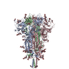
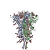
- PDB-7y6d: Cryo-EM structure of SARS-CoV-2 Delta variant spike proteins on i... -
+ Open data

- Basic information
| Entry | Database: PDB / ID: 7y6d | ||||||
|---|---|---|---|---|---|---|---|
| Title | Cryo-EM structure of SARS-CoV-2 Delta variant spike proteins on intact virions: 3 Closed RBD | ||||||
 Components Components | Spike glycoprotein | ||||||
 Keywords Keywords | VIRAL PROTEIN / trimer | ||||||
| Function / homology |  Function and homology information Function and homology informationsymbiont-mediated disruption of host tissue / Maturation of spike protein / Translation of Structural Proteins / Virion Assembly and Release / host cell surface / host extracellular space / symbiont-mediated-mediated suppression of host tetherin activity / Induction of Cell-Cell Fusion / structural constituent of virion / membrane fusion ...symbiont-mediated disruption of host tissue / Maturation of spike protein / Translation of Structural Proteins / Virion Assembly and Release / host cell surface / host extracellular space / symbiont-mediated-mediated suppression of host tetherin activity / Induction of Cell-Cell Fusion / structural constituent of virion / membrane fusion / entry receptor-mediated virion attachment to host cell / Attachment and Entry / host cell endoplasmic reticulum-Golgi intermediate compartment membrane / positive regulation of viral entry into host cell / receptor-mediated virion attachment to host cell / host cell surface receptor binding / symbiont-mediated suppression of host innate immune response / endocytosis involved in viral entry into host cell / receptor ligand activity / fusion of virus membrane with host plasma membrane / fusion of virus membrane with host endosome membrane / viral envelope / symbiont entry into host cell / virion attachment to host cell / SARS-CoV-2 activates/modulates innate and adaptive immune responses / host cell plasma membrane / virion membrane / identical protein binding / membrane / plasma membrane Similarity search - Function | ||||||
| Biological species |   Severe acute respiratory syndrome coronavirus 2 Severe acute respiratory syndrome coronavirus 2 | ||||||
| Method | ELECTRON MICROSCOPY / single particle reconstruction / cryo EM / Resolution: 4.39 Å | ||||||
 Authors Authors | Xu, J. / Song, Y. / Li, S. | ||||||
| Funding support |  China, 1items China, 1items
| ||||||
 Citation Citation | Journal: Proc Natl Acad Sci U S A / Year: 2023 Title: In situ architecture and membrane fusion of SARS-CoV-2 Delta variant. Authors: Yutong Song / Hangping Yao / Nanping Wu / Jialu Xu / Zheyuan Zhang / Cheng Peng / Shibo Li / Weizheng Kong / Yong Chen / Miaojin Zhu / Jiaqi Wang / Danrong Shi / Chongchong Zhao / Xiangyun ...Authors: Yutong Song / Hangping Yao / Nanping Wu / Jialu Xu / Zheyuan Zhang / Cheng Peng / Shibo Li / Weizheng Kong / Yong Chen / Miaojin Zhu / Jiaqi Wang / Danrong Shi / Chongchong Zhao / Xiangyun Lu / Martín Echavarría Galindo / Sai Li / Abstract: Among the current five Variants of Concern, infections caused by SARS-CoV-2 B.1.617.2 (Delta) variant are often associated with the greatest severity. Despite recent advances on the molecular basis ...Among the current five Variants of Concern, infections caused by SARS-CoV-2 B.1.617.2 (Delta) variant are often associated with the greatest severity. Despite recent advances on the molecular basis of elevated pathogenicity using recombinant proteins, the architecture of intact Delta virions remains veiled. Moreover, pieces of molecular evidence for the detailed mechanism of S-mediated membrane fusion are missing. Here, we showed the pleomorphic nature of Delta virions from electron beam inactivated samples and reported the in situ structure and distribution of S on the authentic Delta variant. We also captured the virus-virus fusion events, which provided pieces of structural evidence for Delta's attenuated dependency on cellular factors for fusion activation, and proposed a model of S-mediated membrane fusion. Besides, site-specific glycan analysis revealed increased oligomannose-type glycosylation of native Delta S than that of the WT S. Together, these results disclose distinctive factors of Delta being the most virulent SARS-CoV-2 variant. | ||||||
| History |
|
- Structure visualization
| Structure viewer | Molecule: MolmilJmol/JSmol |
|---|
- Downloads & links
-Download
| PDBx/mmCIF format | 7y6d.cif.gz | 563.8 KB | Display | PDBx/mmCIF format |
|---|---|---|---|---|
| PDB format | pdb7y6d.ent.gz | 452.6 KB | Display | PDB format |
| PDBx/mmJSON format | 7y6d.json.gz | Tree view | PDBx/mmJSON format | |
| Others |  Other downloads Other downloads |
-Validation report
| Arichive directory | https://data.pdbj.org/pub/pdb/validation_reports/y6/7y6dftp://data.pdbj.org/pub/pdb/validation_reports/y6/7y6d | HTTPS FTP |
|---|
-Related structure data
| Related structure data | 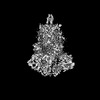 33205MC M: map data used to model this data C: citing same article ( |
|---|---|
| Similar structure data |


-Links
 PDBj
PDBj


- Assembly
| Deposited unit | 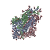
|
|---|---|
| 1 |
|
-Components
| #1: Protein | Mass: 141484.719 Da / Num. of mol.: 3 / Source method: isolated from a natural source Details: T19R, G142D, L452R, T478K, D614G, P681R, D950N are Variants observed in Delta. Source: (natural) Severe acute respiratory syndrome coronavirus 2Variant: Delta variant / References: UniProt: P0DTC2 #2: Polysaccharide | 2-acetamido-2-deoxy-beta-D-glucopyranose-(1-4)-2-acetamido-2-deoxy-beta-D-glucopyranose Source method: isolated from a genetically manipulated source #3: Sugar | ChemComp-NAG /   Type: D-saccharide, beta linking / Mass: 221.208 Da / Num. of mol.: 33 Type: D-saccharide, beta linking / Mass: 221.208 Da / Num. of mol.: 33Source method: isolated from a genetically manipulated source Formula: C8H15NO6 Has ligand of interest | N | Has protein modification | Y | |
|---|
-Experimental details
-Experiment
| Experiment | Method: ELECTRON MICROSCOPY |
|---|---|
| EM experiment | Aggregation state: PARTICLE / 3D reconstruction method: single particle reconstruction |
- Sample preparation
| Component | Name: Severe acute respiratory syndrome coronavirus 2 / Type: VIRUS / Entity ID: #1 / Source: NATURAL | |||||||||||||||||||||||||
|---|---|---|---|---|---|---|---|---|---|---|---|---|---|---|---|---|---|---|---|---|---|---|---|---|---|---|
| Molecular weight | Experimental value: NO | |||||||||||||||||||||||||
| Source (natural) | Organism: Severe acute respiratory syndrome coronavirus 2 | |||||||||||||||||||||||||
| Details of virus | Empty: NO / Enveloped: YES / Isolate: STRAIN / Type: VIRION | |||||||||||||||||||||||||
| Natural host | Organism: Homo sapiens | |||||||||||||||||||||||||
| Buffer solution | pH: 7.4 | |||||||||||||||||||||||||
| Buffer component |
| |||||||||||||||||||||||||
| Specimen | Embedding applied: NO / Shadowing applied: NO / Staining applied: NO / Vitrification applied: YES | |||||||||||||||||||||||||
| Specimen support | Grid material: COPPER / Grid type: Quantifoil R1.2/1.3 | |||||||||||||||||||||||||
| Vitrification | Instrument: GATAN CRYOPLUNGE 3 / Cryogen name: NITROGEN |
- Electron microscopy imaging
| Experimental equipment |  Model: Titan Krios / Image courtesy: FEI Company |
|---|---|
| Microscopy | Model: FEI TITAN KRIOS |
| Electron gun | Electron source:  FIELD EMISSION GUN / Accelerating voltage: 300 kV / Illumination mode: FLOOD BEAM FIELD EMISSION GUN / Accelerating voltage: 300 kV / Illumination mode: FLOOD BEAM |
| Electron lens | Mode: BRIGHT FIELD / Nominal magnification: 81000 X / Nominal defocus max: 3000 nm / Nominal defocus min: 1000 nm / Cs: 2.7 mm |
| Specimen holder | Cryogen: NITROGEN |
| Image recording | Electron dose: 50 e/Å2 / Film or detector model: GATAN K3 (6k x 4k) / Num. of real images: 25851 |
| Image scans | Width: 5760 / Height: 4092 |
- Processing
| EM software |
| ||||||||||||||||||||||||||||||||
|---|---|---|---|---|---|---|---|---|---|---|---|---|---|---|---|---|---|---|---|---|---|---|---|---|---|---|---|---|---|---|---|---|---|
| CTF correction | Details: CTFs are applied to the projections of the map during 3D classification and 3D refinement. Type: PHASE FLIPPING AND AMPLITUDE CORRECTION | ||||||||||||||||||||||||||||||||
| Particle selection | Num. of particles selected: 674792 Details: Initial micrographs were deconvolved using Warp, and 7,950 spikes were manually picked from 474 micrographs for training Topaz neural network. The trained Topaz neural network was used to ...Details: Initial micrographs were deconvolved using Warp, and 7,950 spikes were manually picked from 474 micrographs for training Topaz neural network. The trained Topaz neural network was used to automatically pick the spikes. 674,792 particles were auto-picked by Topaz. | ||||||||||||||||||||||||||||||||
| Symmetry | Point symmetry: C3 (3 fold cyclic) | ||||||||||||||||||||||||||||||||
| 3D reconstruction | Resolution: 4.39 Å / Resolution method: FSC 0.143 CUT-OFF / Num. of particles: 45252 / Algorithm: BACK PROJECTION / Num. of class averages: 1 / Symmetry type: POINT | ||||||||||||||||||||||||||||||||
| Atomic model building | B value: 118.79 / Space: REAL | ||||||||||||||||||||||||||||||||
| Atomic model building | 3D fitting-ID: 1 / Pdb chain-ID: A / Source name: PDB / Type: experimental model
|